Associate Professor of Pediatrics Chang Gung Memorial Hospital, Linkou Taipei, Taipei, Taiwan (Republic of China)
Background: Intraventricular hemorrhage (IVH) is a severe complication that adversely affects cerebral hemodynamics in extremely preterm infants. Objective: To investigate minute-by-minute continuous hemodynamic parameters to distinguish between extremely preterm infants with and without IVH. Design/Methods: This post-hoc analysis is based on a prospective observational study involving preterm infants with a gestational age (GA) ≤28 weeks or a birthweight (BW) ≤1000 g. Minute-by-minute hemodynamic data were collected over the first 72 hours, including regional cerebral saturation (rcSO₂) measured by near-infrared spectroscopy, plus cardiac output (CO), arterial saturation (Sat) and perfusion index (PI) using electrical cardiometry with Masimo pulse oximetry. The fraction of tissue oxygen extraction (FTOE) was calculated accordingly. Each infant underwent daily brain ultrasounds during the first week to screen for IVH. Hemodynamic parameters were compared between the IVH and non-IVH groups using the mean and coefficient of variation (CV), as well as demographics, inotrope usage, and the presence of significant patent ductus arteriosus (sPDA, diameter ≥2 mm, left atrium/aortic root ≥1.5). Parameters were averaged every 6 hours and assessed with repeated-measures analysis of variance (RM-ANOVA). Cerebral autoregulation was evaluated using rolling correlations (30-minute window) for CO–rcSO₂ and CO–FTOE, and correlation degrees were compared. Study protocol is illustrated in Fig 1. Results: A total of 30 extremely preterm infants (GA 27.1±2.0 weeks, BW 830±225 g) were enrolled; 6 were diagnosed with IVH (1 grade 4, and 5 grade 1–2) in the first week. As shown in Table 1, there were no difference in demographics, inotrope, sPDA, CO, Sat or PI. Although the IVH group showed only marginally lower overall rcSO2 (73.9±7.6 vs. 78.9±4.8 %, p=0.054) and higher FTOE (21.2±6.4 vs. 17.1±3.9 %, p=0.052), they exhibited a significantly greater CV of rcSO2 (11.6±3.4 vs. 8.2±2.6, p=0.008), indicating greater cerebral oxygenation variability. A trend towards lower rcSO₂ was observed in the IVH group (between-group RM-ANOVA p=0.037), particularly after 42 hours of life (Fig 2). The IVH group showed more frequent high correlations (r>0.7) in CO–rcSO₂ and less frequent low correlations (r=0.4– -0.7) in CO–FTOE, suggesting impaired cerebral autoregulation.
Conclusion(s): Continuous hemodynamic monitoring revealed that infants with IVH experienced greater rcSO₂ fluctuations, a trend toward lower rcSO₂, and impaired autoregulation. These findings highlight the potential for real-time monitoring to identify IVH risk in extremely preterm infants.
Fig 1. Study protocol 
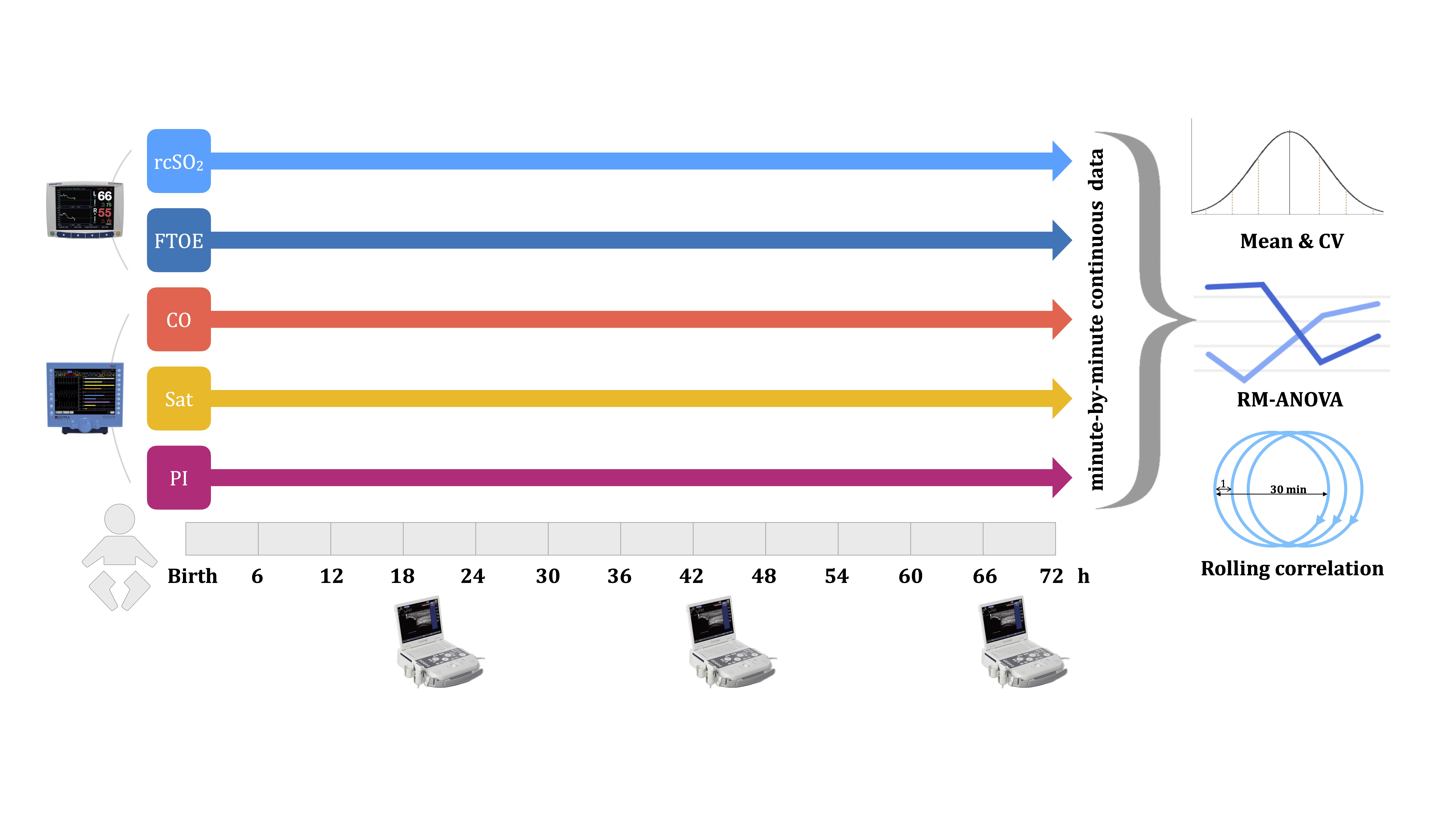
Hemodynamic parameters, including regional cerebral saturation (rcSO₂) , fraction of tissue oxygen extraction (FTOE), cardiac output (CO), arterial saturation (Sat) and perfusion index (PI), were collected minute-by-minute over the first 72 hours. Minute-by-minute data were analyzed between the IVH and non-IVH groups using the mean and coefficient of variation (CV), repeated-measures analysis of variance (RM-ANOVA), and rolling correlations.
Table 1. Demographic and hemodynamic characteristics between extremely preterm infants with and without IVH. 
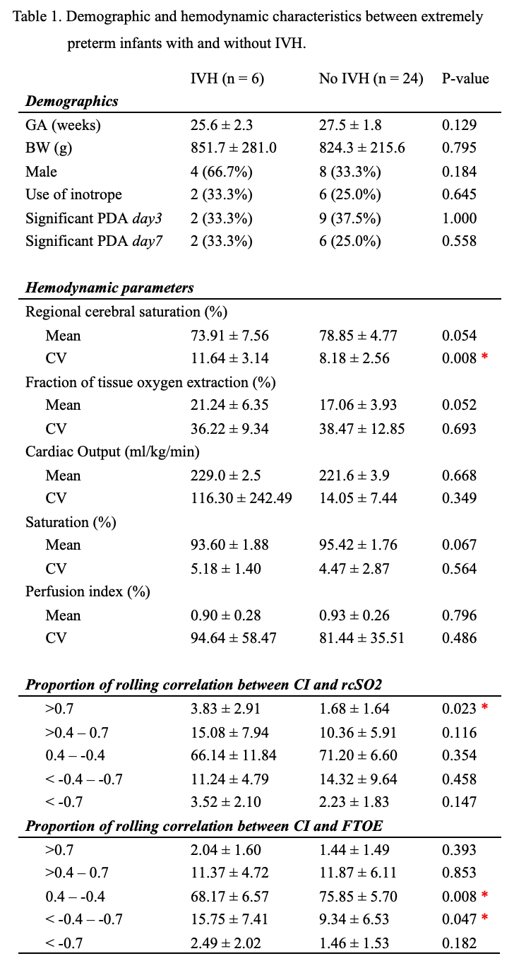
The IVH group exhibited a significantly greater CV of rcSO₂ , more frequent high correlations (r>0.7) in CO–rcSO₂ and less frequent low correlations (r=0.4– -0.7) in CO–FTOE. * indicates statistic significance (p <0.05).
Fig 2. The trend of rcSO₂ between extremely preterm infants with and without IVH. 
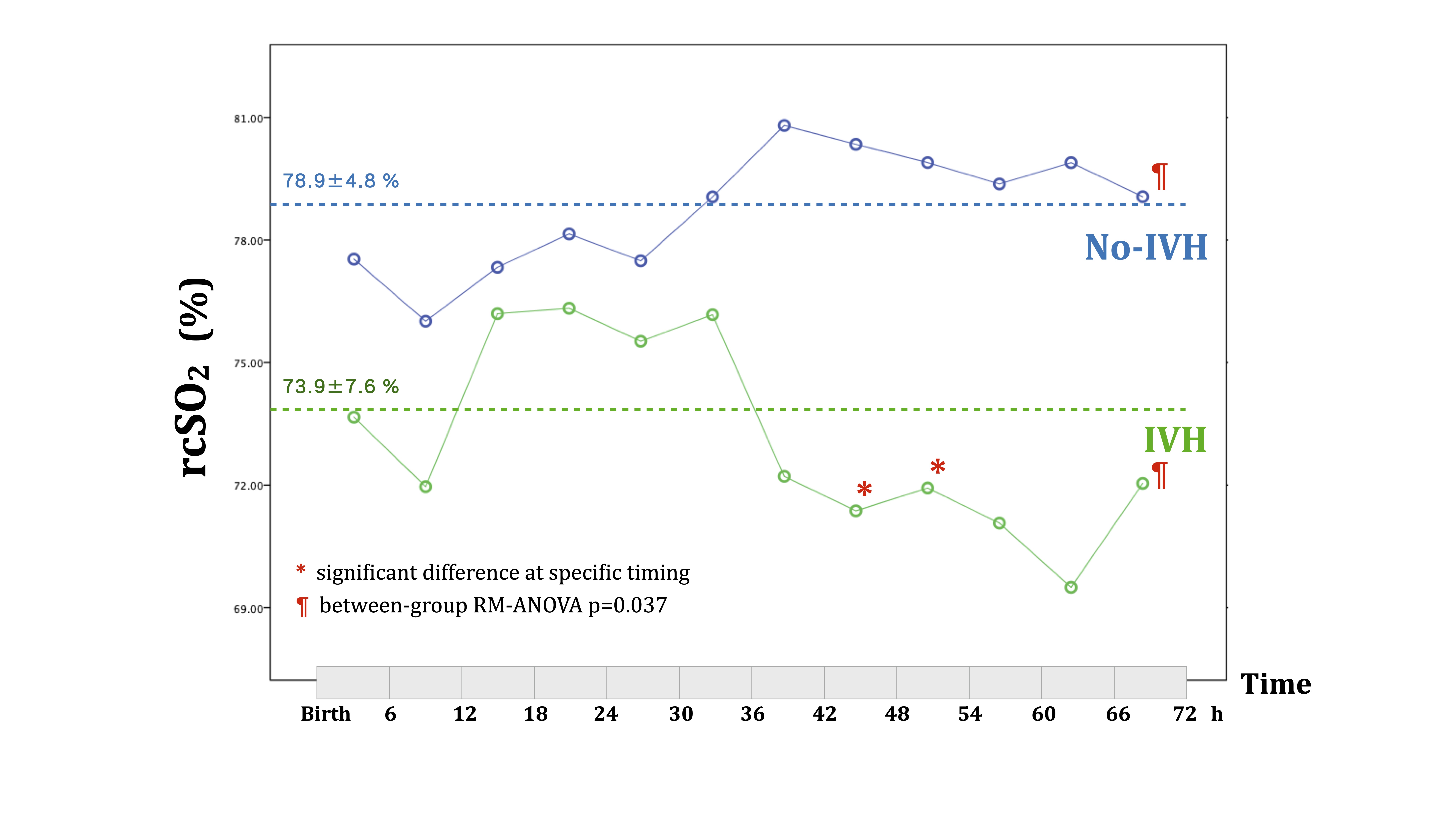
A trend towards lower rcSO₂ was observed in the IVH group (¶ between-group RM-ANOVA p=0.037), particularly after 42 hours of life (*).