
Neonatal General 2: Neurology
Session: Neonatal General 2: Neurology
Lynn Bitar, MD MSc (she/her/hers)
Pediatric Resident
University of Texas Southwestern Medical School
Dallas, Texas, United States
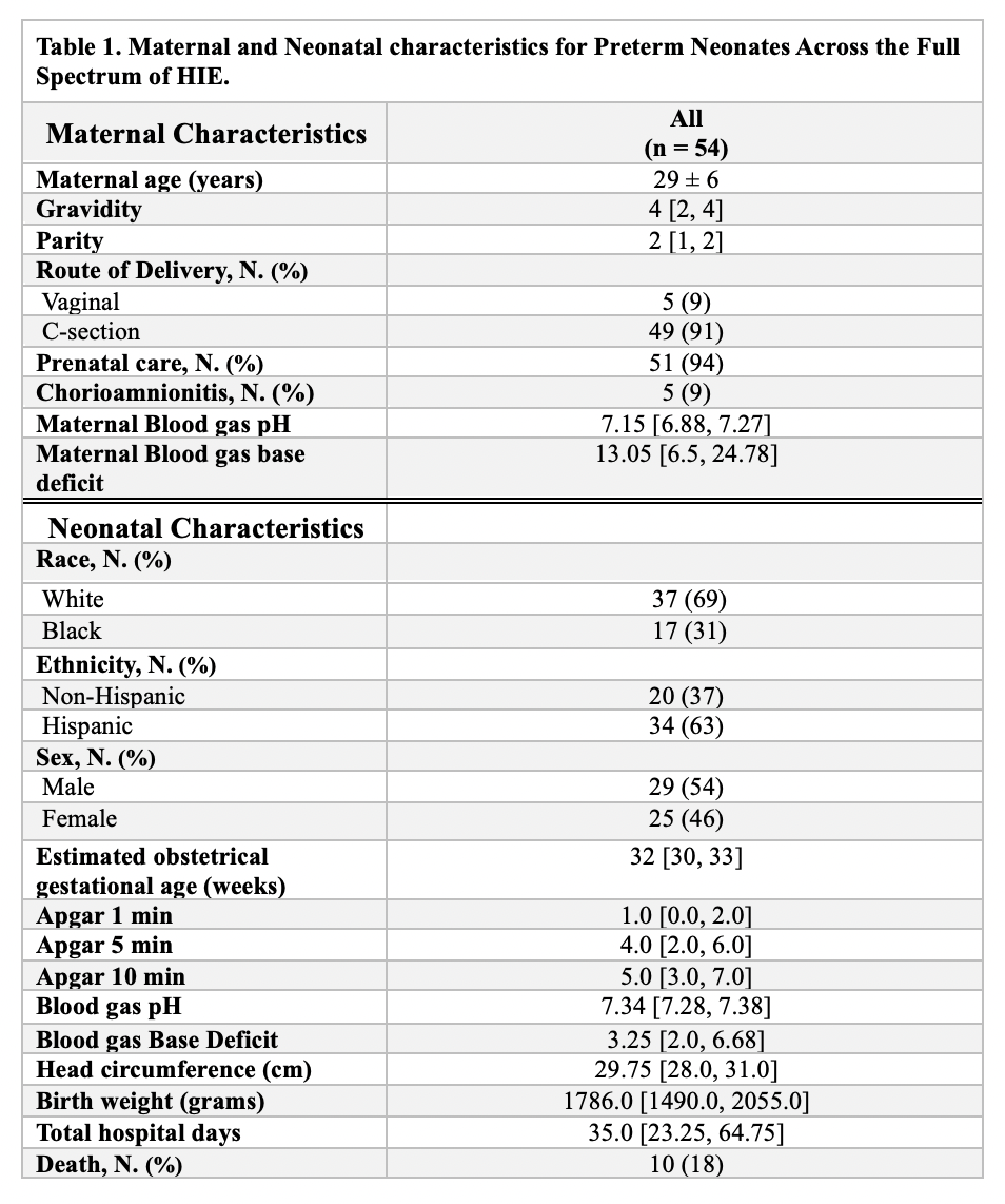 For continuous variables, normality was tested using the Shapiro-Wilk test. Variables with a normal distribution are reported with the mean and standard deviation, while non-normal distributions are summarized with the median and the 25th and 75th percentiles. Categorical variables are presented with counts (N) and percentages (%).
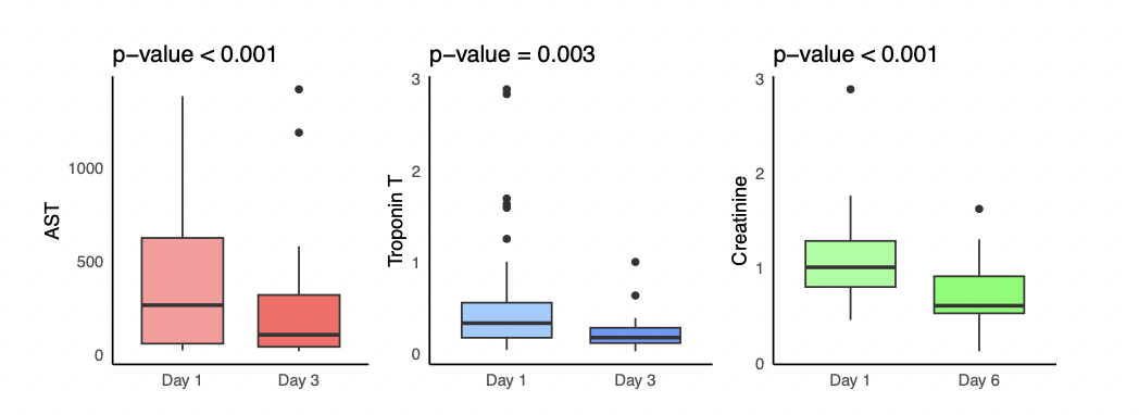 Distribution of AST, Troponin T, and Creatinine showing significant differences between Day 1 and Day 3, as determined by the paired Wilcoxon rank-sum test.For continuous variables, normality was tested using the Shapiro-Wilk test. Variables with a normal distribution are reported with the mean and standard deviation, while non-normal distributions are summarized with the median and the 25th and 75th percentiles. Categorical variables are presented with counts (N) and percentages (%).Distribution of AST, Troponin T, and Creatinine showing significant differences between Day 1 and Day 3, as determined by the paired Wilcoxon rank-sum test.