Neonatal Neurology 2
Session: Neonatal Neurology 2
353 - Major microstructural alterations in neural connectivity associated with moderate prematurity, observed without substantial volumetric differences
Friday, April 25, 2025
5:30pm - 7:45pm HST
Publication Number: 353.5996
Océane-Rose Leloutre-Salat, Universite de Montreal Faculty of Medicine, Laval, PQ, Canada; Ophélia Dalkiriadis, Universite de Montreal Faculty of Medicine, Montréal, PQ, Canada; Erjun Zhang, Polytechnique Montreal, Montreal, PQ, Canada; Farzad Beizaee, CHU Sainte-Justine, Montreal, PQ, Canada; Natacha Paquette, Sainte-Justine Hospital Research Center, Montréal, PQ, Canada; Elana Pinchefsky, CHU Sainte-Justine, Montreal, PQ, Canada; Anne Gallagher, University of Montreal, Montreal, PQ, Canada; Gregory Lodygensky, University of Montreal, Montreal, PQ, Canada
- OL
Océane-Rose Leloutre-Salat, 20220063 (she/her/hers)
Student
Universite de Montreal Faculty of Medicine
Laval, Quebec, Canada
Presenting Author(s)
Background: This study examines volumetric and connectivity alterations associated with prematurity, with a focus on limbic system integrity in moderate preterm infants. In 2020, 9.9% of births worldwide were preterm , and although mortality rates have decreased, neurodevelopmental morbidity remains a significant contributor to cognitive and language delays. In preterm children, distinct volumetric alterations are observed in the right hippocampus.
Objective: We hypothesize that preterm infants exhibit impaired connectivity in the fornix and uncinate fasciculus, as well as reduced volumes in key limbic structures, including the amygdala, hippocampus, and frontal lobe.
Design/Methods: The study conducted T2-weighted MRI on a 3T Siemens MAGNETOM Skyra scanner in a cohort of 37 participants, including 18 healthy full-term newborns and 19 preterm newborns. A U-Net convolutional neural network with 98 tissue classes, trained specifically on neonatal data, was employed to segment the amygdala, hippocampus, and frontal lobe. Diffusion tensor imaging (DTI) in DSI-Studio was then used to evaluate the integrity of tracts originating from these structures, focusing on the fornix and uncinate fasciculus. To ensure reproducibility of this neonatal DSI-Studio protocol, inter- and intra-rater reliability was assessed with spatial matching analysis by κ.
Results: Volume analyses were performed on 30 MRI scans, including 13 full-term newborn controls and 17 preterm infants. No significant volumetric alterations were detected in the right and left hippocampus between the control group and the preterm group (p-values = 0.4570 and 0.6131). Similarly, no significant volumetric alterations were observed in the right and left amygdala between the two groups (p-values = 0.3851 and 0.7964).
Diffusion tensor imaging (DTI) metrics were analyzed for 37 participants, including 18 full-term newborns and 19 preterm infants. Significant alterations were observed in the radial diffusivity (RD) of the right and left fornix between the two groups (p-values = 0.0002 and < 0.0001). Significant connectivity differences were also detected in the uncinate fasciculus for the control group and the preterm group (p-values = 0.0011 and < 0.0001).
Conclusion(s): This study reveals major limbic connectivity impairment in preterm newborns, despite preserved volumetric integrity of associated structures. These findings highlight how detailed microstructural analyses can uncover significant changes that might otherwise go undetected. The implications of these results are substantial, suggesting potential concerns for neonatal neurodevelopment.
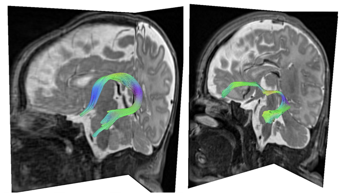
Figure 1. Acquisition of the fornix and the uncinate fasciculus in a newborn, displayed on a color map from DSI-Studio.
 The image was obtained following a DTI performed on a 3.0 T Siemens scanner, equipped with 98 tissue classes, a slice thickness of 2 mm, TE of 81 ms, TR of 8,000 ms, FOV of 220 mm x 220 mm, matrix size of 220 mm x 220 mm, and a b-value of 700 s/mm². The fractional anisotropy and angular thresholds were set to 0.12 and 60 degrees, respectively.
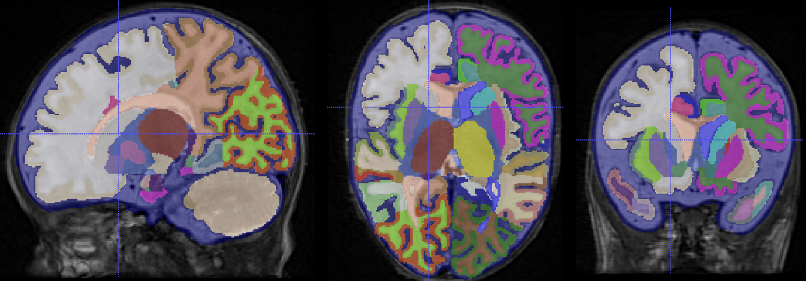
Figure 2. Segmentation on ITK-SNAP of 98 tissue classes using the MCRIB-35 convolutional neural network.
 Performed on a T2-weighted MRI with a 3.0 T Siemens scanner, featuring a slice thickness of 2 mm, matrix size of 192 mm x 192 mm, TR = 12,020 ms, TE = 90 ms, and FOV = 120 mm x 120 mm, on a control subject. The sagittal (A), axial (B), and coronal (C) slices highlight structures such as the hippocampus and the amygdala in both the right and left hemispheres, which can subsequently be extracted as voxels or volume data.
Table 1. Data on gestational age and age at MRI for the control newborns (n=13) and preterm infants (n=17) as well as the median and standard deviation of hippocampal and amygdala volumes, along with radial diffusivity (RD) of the fornix and uncinate fasciculus.
.png) The gestational age shows a significant difference between groups, with a p-value of < 0.0001. Gestational age at MRI does not show a significant difference, with a p-value of 0.0540. No significant differences were found in the volumes of the left and right hippocampus (p = 0.6131and 0.4570) or in the amygdala volume (p = 0.3851 and 0.7964). Significant differences were observed between groups for the right and left fornix (p = 0.0002 and < 0.0001) as well as for the uncinate fasciculus (p = 0.0011 and < 0.0001).
The gestational age shows a significant difference between groups, with a p-value of < 0.0001. Gestational age at MRI does not show a significant difference, with a p-value of 0.0540. No significant differences were found in the volumes of the left and right hippocampus (p = 0.6131and 0.4570) or in the amygdala volume (p = 0.3851 and 0.7964). Significant differences were observed between groups for the right and left fornix (p = 0.0002 and < 0.0001) as well as for the uncinate fasciculus (p = 0.0011 and < 0.0001).
