
Neonatal Neurology 2
Session: Neonatal Neurology 2
Océane-Rose Leloutre-Salat, 20220063 (she/her/hers)
Student
Universite de Montreal Faculty of Medicine
Laval, Quebec, Canada
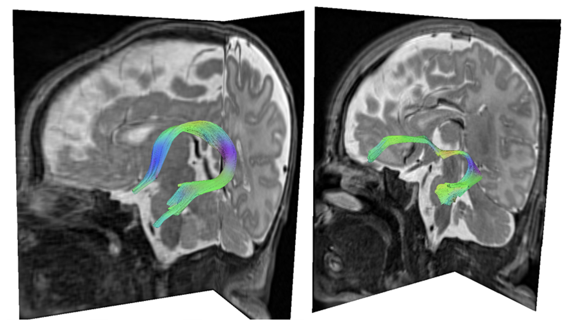 The image was obtained following a DTI performed on a 3.0 T Siemens scanner, equipped with 98 tissue classes, a slice thickness of 2 mm, TE of 81 ms, TR of 8,000 ms, FOV of 220 mm x 220 mm, matrix size of 220 mm x 220 mm, and a b-value of 700 s/mm². The fractional anisotropy and angular thresholds were set to 0.12 and 60 degrees, respectively.
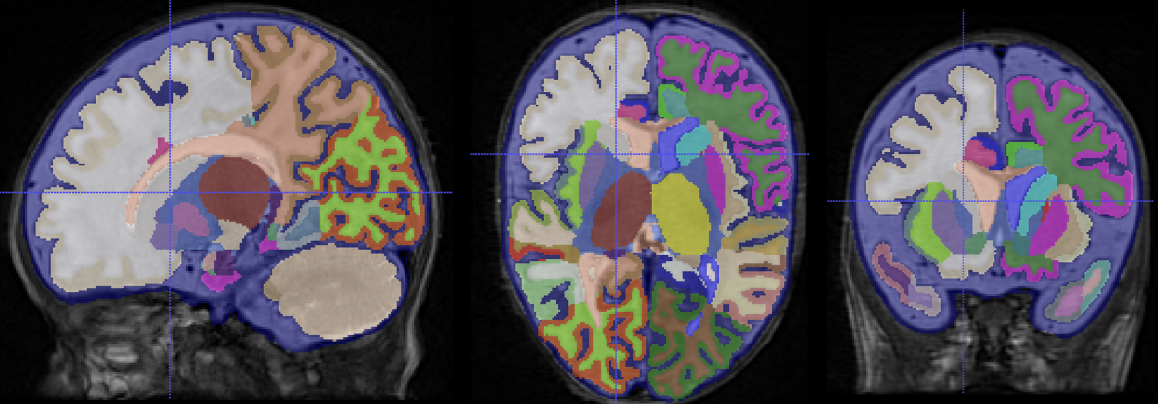 Performed on a T2-weighted MRI with a 3.0 T Siemens scanner, featuring a slice thickness of 2 mm, matrix size of 192 mm x 192 mm, TR = 12,020 ms, TE = 90 ms, and FOV = 120 mm x 120 mm, on a control subject. The sagittal (A), axial (B), and coronal (C) slices highlight structures such as the hippocampus and the amygdala in both the right and left hemispheres, which can subsequently be extracted as voxels or volume data.
.png) The gestational age shows a significant difference between groups, with a p-value of < 0.0001. Gestational age at MRI does not show a significant difference, with a p-value of 0.0540. No significant differences were found in the volumes of the left and right hippocampus (p = 0.6131and 0.4570) or in the amygdala volume (p = 0.3851 and 0.7964). Significant differences were observed between groups for the right and left fornix (p = 0.0002 and < 0.0001) as well as for the uncinate fasciculus (p = 0.0011 and < 0.0001).
The gestational age shows a significant difference between groups, with a p-value of < 0.0001. Gestational age at MRI does not show a significant difference, with a p-value of 0.0540. No significant differences were found in the volumes of the left and right hippocampus (p = 0.6131and 0.4570) or in the amygdala volume (p = 0.3851 and 0.7964). Significant differences were observed between groups for the right and left fornix (p = 0.0002 and < 0.0001) as well as for the uncinate fasciculus (p = 0.0011 and < 0.0001).