
Developmental Biology/Cardiac & Pulmonary Development
Session: Developmental Biology/Cardiac & Pulmonary Development
 photo")
Rachel L. Leon, MD, PhD (she/her/hers)
Assistant Professor
University of Texas Southwestern Medical School
Dallas, Texas, United States
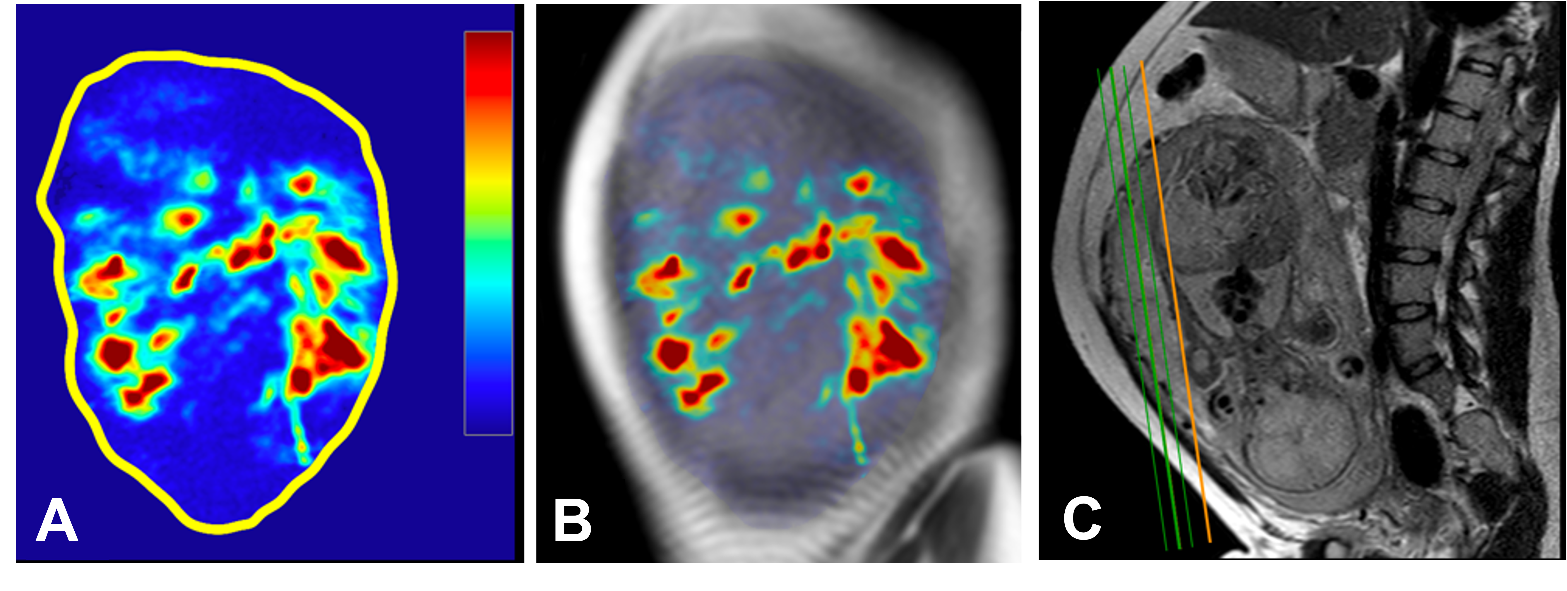 Representative quantitative perfusion map (A), overlaid on anatomical T2-weighted coronal image (B), and the prescribed slices (orange and green lines) shown on sagittal T2 image (C) in a 23 year old pregnant subject with CHD fetus at 32+5 weeks gestation. Colorbar represents 0-300 mL/100g/min.
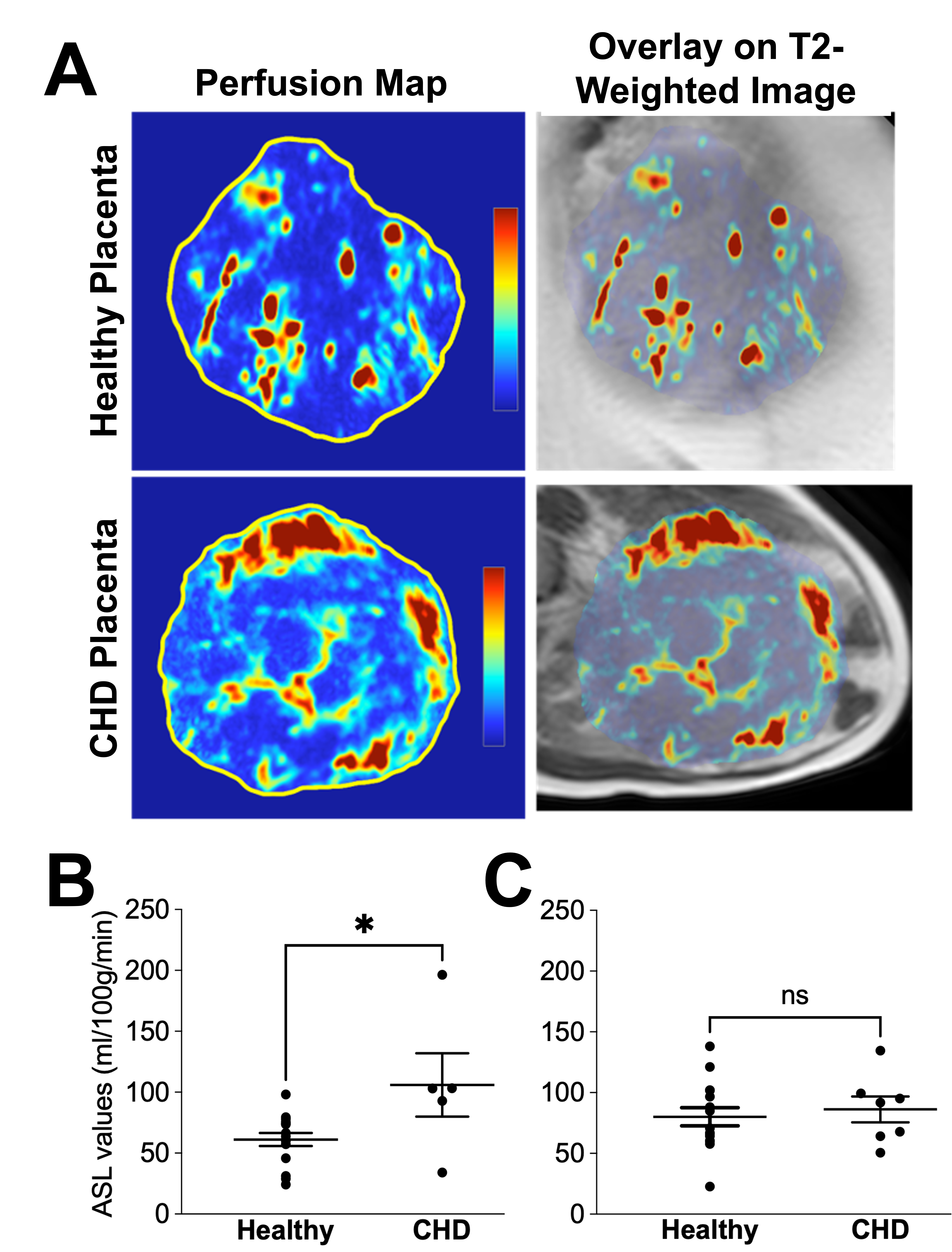 Panel A: Representative quantitative perfusion maps (left) and overlaid perfusion maps on anatomical T2-weighted images (right), shown for a 33 year old pregnant subject with healthy fetus at 28+5 weeks gestation (top) and 35 year old pregnant subject with CHD fetus (Tetrology of Fallot) at 29+2 weeks gestation (bottom). Colorbar represents 0-300 mL/100g/min. Panel B: Quantitative placental perfusion measured using FAIR-ASL showed significantly higher perfusion in CHD fetus compared to healthy fetus at gestation age <32 weeks (P <0.05). Panel C: Placental perfusion values were not significantly different between CHD and healthy fetuses at ≥32 weeks gestation.