
Pulmonology
Session: Pulmonology
 photo")
Chintan K. Gandhi, MBBS, MD (he/him/his)
Associate Professor
Penn State Hershey College of Medicine
Hershey, Pennsylvania, United States
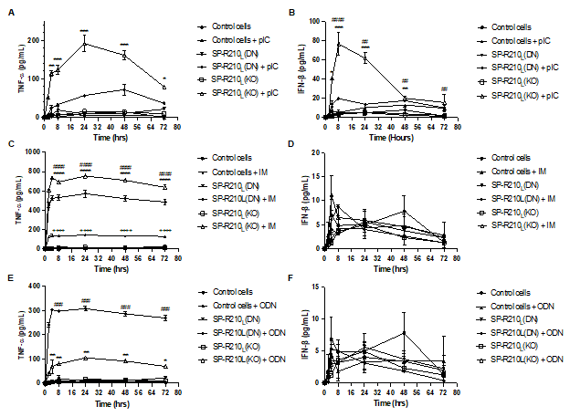 (A) pIC significantly increased TNF-⍺ production in SP-R210(KO) cells (B) For IFN-β secretion, pIC-treated SP-R210(KO) cells showed the strongest response. pIC-treated SP-R210(DN) cells had a lower stimulation of IFN-β secretion compared to SP-R210(KO), yet there was a significant difference compared to untreated cells. (C) IM significantly elevated TNF-⍺ production across all three cell lines with the highest level in SP-R210(KO) cells, followed by SP-R210(DN) and control cells. (D) No significant difference was observed in IFN-β secretion across all three cell lines compared to controls. (E) In contrast to pIC and IM, ODN 1585 elicited higher TNF-α production in SP-R210(DN) cells compared to SP-R210(KO) cells. (F) No significant difference was observed in IFN-β secretion across all three cell lines upon ODN1585 treatment. p < 0.05 between treated versus untreated cells (One-way ANOVA).
 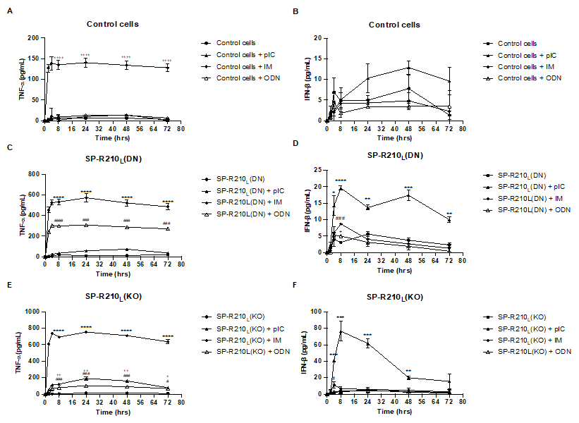 (A) IM was the only treatment that stimulated TNF-⍺ secretion in control cells. (B) No significant difference was observed in IFN-β secretion in control cells across treatments. (C) Both IM and ODN treatment resulted in robust stimulation of TNF-⍺ secretion in SP-R210L(DN) cells compared to untreated cells. However, no significant difference is observed between pIC-treated versus untreated SP-R210L(DN) cells. (D) All three treatments resulted in significant IFN-β secretion in SP-R210L(DN) cells at t = 8h. At later time points, only SP-R210L(DN) cells treated with pIC had a significantly higher IFN-β secretion than untreated cells. (E) All three treatments resulted in significant TNF-⍺ secretion in SP-R210(KO) cells, with IM causing the most robust stimulation, followed by pIC, then ODN. (F) At t = 8-72h, only IM caused the most robust IFN-β stimulation in KO cells. p < 0.05 between treated versus untreated cells (One-way ANOVA).