
Infectious Diseases 2: Bacterial infections
Session: Infectious Diseases 2: Bacterial infections
 photo")
Mit Shah, MD (he/him/his)
PGY3 Med-Peds Resident
McGovern Medical School at the University of Texas Health Science Center at Houston
Houston, Texas, United States
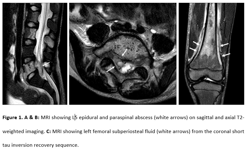 Figure 1. A & B: MRI showing L5 epidural and paraspinal abscess (white arrows) on sagittal and axial T2-weighted imaging. C: MRI showing left femoral subperiosteal fluid (white arrows) from the coronal short tau inversion recovery sequence.
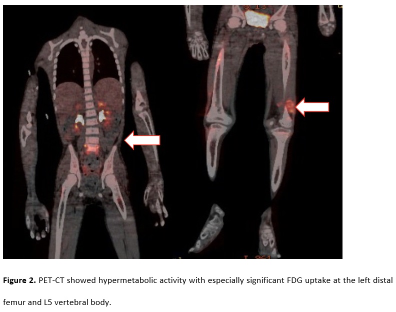 Figure 2. PET-CT showed hypermetabolic activity with especially significant FDG uptake at the left distal femur and L5 vertebral body.
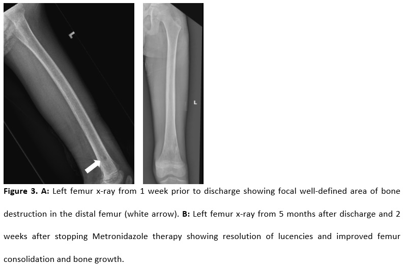 Figure 3. A: Left femur x-ray from 1 week prior to discharge showing focal well-defined area of bone destruction in the distal femur (white arrow). B: Left femur x-ray from 5 months after discharge and 2 weeks after stopping Metronidazole therapy showing resolution of lucencies and improved femur consolidation and bone growth.