
Neonatal Nephrology/AKI 1
Session: Neonatal Nephrology/AKI 1
Sage Timberline, MD
Fellow
University of Virginia
Charlottesville, Virginia, United States
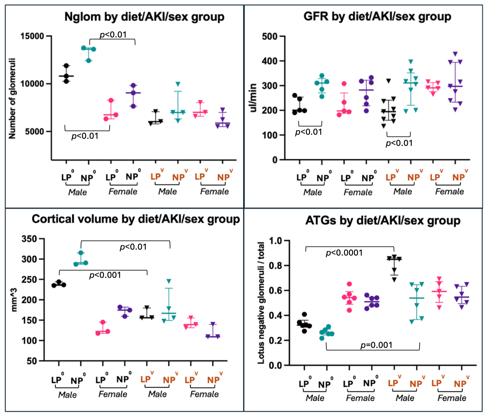 Among control animals, males have significantly more glomeruli than females. In control and AKI males, GFR is lower in the LP group. After AKI, LP and NP males have less cortical volume and more ATG.
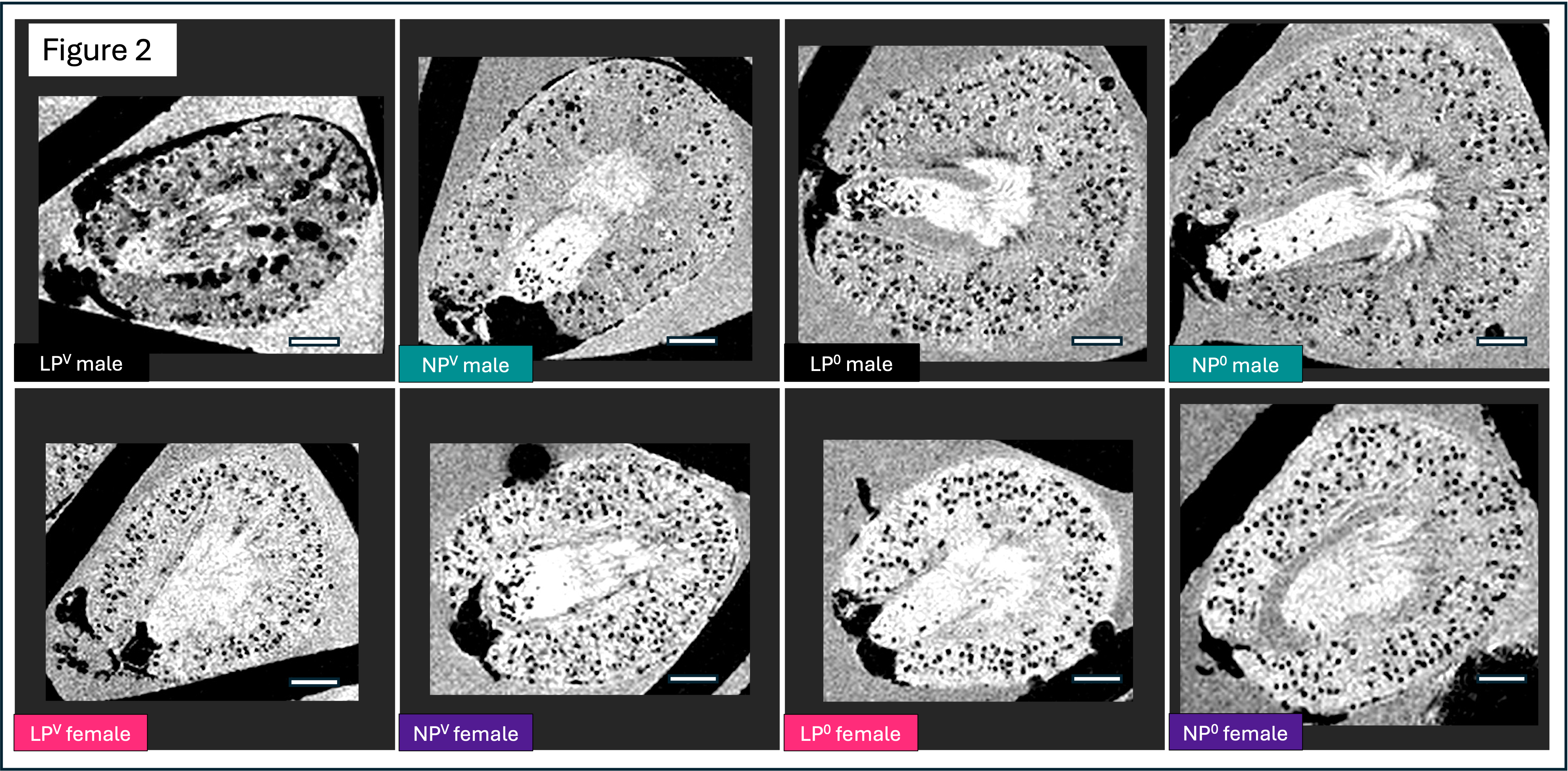 MRIs of LPV males have a pathologic phenotype with abnormally dilated, cationic ferritin-labeled structures. LPV and NPV males have visibly fewer nephrons than LP0 and NP0 males, whereas all female MRIs have a similar appearance. Among control animals, males have significantly more glomeruli than females. In control and AKI males, GFR is lower in the LP group. After AKI, LP and NP males have less cortical volume and more ATG.MRIs of LPV males have a pathologic phenotype with abnormally dilated, cationic ferritin-labeled structures. LPV and NPV males have visibly fewer nephrons than LP0 and NP0 males, whereas all female MRIs have a similar appearance.