602 - Impact of high maternal body mass index on fetal cerebral cortical and cerebellar volumes
Saturday, April 26, 2025
2:30pm – 4:45pm HST
Publication Number: 602.5178
Emiko Takeoka, Hoyogo Prefectural Kobe Children's Hospital, Kobe, Hyogo, Japan; April A. Carlson, University of California, Irvine, School of Medicine, Orange, CA, United States; Neel Madan, Harvard Medical School, Boston, MA, United States; Afshin Azimirad, Tufts University School of Medicine, Boston, MA, United States; Taysir Mahmoud, Tufts Childrens Hospital, Boston, MA, United States; Rie Kitano, Tsuchiura kyodo general hospital, Tsuchiura, Ibaraki, Japan; Shizuko Akiyama, Tohoku University Hospital, Sendai, Miyagi, Japan; HyukJin Yun, Childrens Mercy Hospital, Kansas City, MO, United States; Richard Tucker, Women & Infants Hospital of Rhode Island, Providence, RI, United States; Kiho Im, Boston Children's Hospital, Boston, MA, United States; Perrie O'Tierney-Ginn, Tufts University School of Medicine, Boston, MA, United States; Tomo Tarui, Hasbro Children's Hospital at Rhode Island Hospital, Providence, RI, United States
Associate Professor of Pediatrics and Neurology Hasbro Children's at Rhode Island Hospital Providence, Rhode Island, United States
Background: Maternal obesity increases a child’s risk of neurodevelopmental impairment. However, little is known about the impact of maternal obesity on fetal brain development. Objective: We aim to quantitatively analyze the impact of maternal obesity on fetal brain growth. Design/Methods: We prospectively recruited 20 healthy pregnant women across the range of pre-pregnancy or first-trimester body mass index (BMI) and performed fetal brain magnetic resonance imaging (MRI) of their healthy singleton fetuses. We examined correlations between early pregnancy maternal BMI and regional brain volume of living fetuses using volumetric MRI analysis. Results: Of 20 fetuses, there were 8 males and 12 females (median gestational age at MRI acquisition was 24.3 weeks, range: 19.7 - 33.3 weeks, median maternal age was 33.3 years, range: 22.0 - 37.4 years). There were no significant differences in clinical demographics between overweight (OW, 25≤BMI < 30)/obese (OB, BMI≥30 kg/m2) (n=12) and normal BMI (18.5≤BMI < 25) (n=8) groups. Fetuses in the OW/OB group had significantly larger left cortical plate (p=0.0003), right cortical plate (p=0.0002), and whole cerebellum (p=0.049) compared to the normal BMI group. In the OW/OB BMI group, cortical plate volume was larger relative to other brain regions after 28 weeks.
Conclusion(s): This pilot study supports the concept that maternal obesity impacts fetal brain volume, detectable via MRI in living fetuses using quantitative analysis.
Maternal obesity accerelates fetal cortical and cerebellar volume growths 
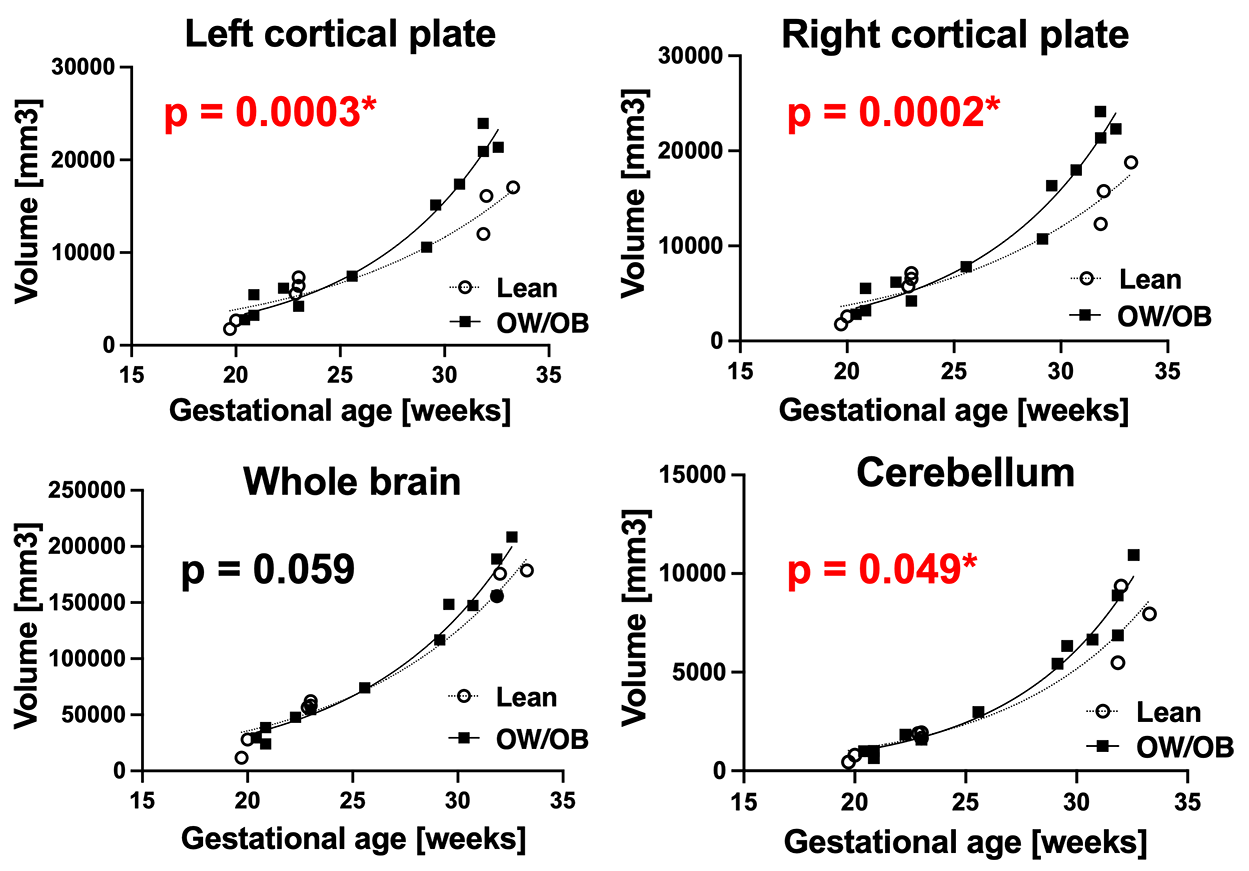
Comparison of regional brain volume between the normal BMI group (○,n=8) and overweight/obesity (OW/OB) group (■,n=12). The fetuses in OW/OB BMI group had significantly larger left and right cortical plates and cerebellum in late pregnancy (after 28 weeks) compared to the normal BMI group.