Assistant professor University of Arkansas for Medical sciences Little Rock, Arkansas, United States
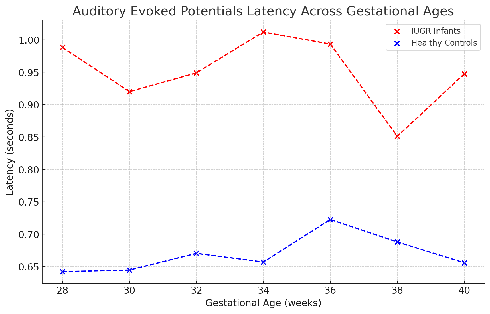
Background: Intrauterine growth restriction (IUGR) affects 5-10% of pregnancies and is linked to 3 to 4 fold increase in mortality and adverse neurodevelopmental (ND) outcomes. Chronic hypoxia in IUGR may disrupt brain development, with studies indicating that up to 30% of affected infants experience lasting cognitive and behavioral impairments. Traditional assessment methods have limitations, while Magnetoencephalography (MEG) provides a non-invasive, real-time measurement of functional brain activity. At the University of Arkansas for Medical Sciences, 15% pregnancies are diagnosed with IUGR, much higher than the national average, highlighting its prevalence and the pressing need for targeted research and intervention strategies at this institution. Objective: To utilize MEG for monitoring brain developmental trajectories in IUGR neonates versus healthy controls to identify and inform on ND delays for targeted therapy. Design/Methods: This controlled, prospective cohort study, monitors 30 infants (15 IUGR and 15 controls). MEG scans are performed at 28-32 weeks gestation and at 1 and 3 months postnatally, focusing on the latency and amplitude of auditory and visual evoked potentials (AER/VER), as well as oscillatory activities across multiple frequency bands. Our study utilizes mixed-model ANOVAs to assess differences in neural activity across time points and between groups, and multivariate regression models to link MEG outcomes to clinical neurological assessments, adjusting for confounders such as gestational age and sex, with a significance threshold set at p< 0.05. Results: Preliminary results from our ongoing study, reveal measurable differences in the ND trajectories in IUGR compared to controls. Initial analyses indicate that infants with IUGR exhibit delayed AERs, with latency times extended by 10-15 milliseconds beyond the norm for healthy infants at similar gestational ages. Furthermore, alterations in resting-state connectivity patterns, such as decreased coherence in the alpha and theta frequency bands, are evident. These early findings, consistent with previous studies, suggest significant disruptions in the maturation of neural activity, which could serve as early indicators for potential cognitive and motor developmental delays.
Conclusion(s): MEG presents a viable tool for enhancing our understanding of the ND impact of IUGR on infants. By providing detailed, non-invasive insights into brain activity, MEG can help pinpoint developmental anomalies at a critical early stage, enabling timely interventions.