665 - The Effects of Fetal Exposure to Cannabis on Neuronal Connectivity and Neuro-Metabolic Profiles in Newborns
Sunday, April 27, 2025
8:30am – 10:45am HST
Publication Number: 665.6974
Ophélia Dalkiriadis, Universite de Montreal Faculty of Medicine, Montréal, PQ, Canada; Océane-Rose Leloutre-Salat, Universite de Montreal Faculty of Medicine, Laval, PQ, Canada; Dinesh K. Deelchand, University of Minnesota Medical School, Minneapolis, MN, United States; Farzad Beizaee, CHU Sainte-Justine, Montreal, PQ, Canada; Jose Dolz, ETS Montreal, Montreal, PQ, Canada; Helge J.. Zöllner, Johns Hopkins University School of Medicine, Baltimore, MD, United States; Gregory Lodygensky, University of Montreal, Montreal, PQ, Canada
Clinical Associate Professor University of Montreal Montreal, Quebec, Canada
Background: Cannabis consumption among pregnant individuals has risen post-legalization, despite concerns about potential neurotoxic impacts on developing fetuses. Objective: This study explores the effects of prenatal cannabis exposure on newborn neurodevelopment, particularly focusing on alterations in neuro-connectivity and neuro-metabolic profiles. The hypothesis posits that cannabis exposure during pregnancy disrupts brain structures abundant in cannabinoid receptors, such as the amygdala, hippocampus, and frontal lobe, leading to connectivity, metabolic and volume abnormalities. Design/Methods: A cohort study recruited 52 participants: 20 cannabis-exposed infants and 32 controls.After postpartum recruitment, neonates part of the subgroup who consented to the MRI underwent resting-state functional MRI (rs-fMRI) and magnetic resonance spectroscopy (MRS) to assess neural connectivity and neuro-metabolic alterations. T2-weighted images were acquired on a 3T Skyra. A trained U-Net convolutional neural network (Beizaee F, Sci Rep. 2023) was used to segment the amygdala and hippocampus. A single-voxel MRS (V=15x 15 x 15 mm3) was positioned on the patient's left amygdala. We also included the acquisition of a water-reference peak. Data analysis was performed using the Osprey toolbox (Oeltzschner et al., 2020). To adapt the software to our experimental conditions, the T1 and T2 relaxation times were corrected for gray matter, white matter, and water (Vanderhasselt et al., 2021). Results: Significant volume reductions were observed in the right amygdala (p = 0.040), and in the left hippocampus (p = 0.011) (table 1). Twenty-five rs-fMRI were analyzed, including 6 cannabis exposed babies and 19 controls. Cannabis-exposed neonates exhibited reduced connectivity in critical brain areas, including right motor and left attention networks, compared to controls. Connectivity differences were prominent between the primary visual area and the right angular gyrus (p=0.038) and between the right parietal region and the frontal pole (p=0.018), indicating impaired synchrony. Spectroscopy results revealed increased myo-inositol levels, indicating inflammation in cannabis-exposed neonates (p < 0.01). Trends toward lower N-acetylaspartate (NAA) and glutamate levels suggest diminished neuronal density in the amygdala.
Conclusion(s): Prenatal cannabis exposure is associated with neuroinflammatory markers and disruptions in functional connectivity in key cognitive and emotional regulatory regions. The findings underline potential long-term neurodevelopmental risks, including cognitive and behavioral challenges.
Concentration of different metabolites in the left amygdala spectro.pdfComparison of the concentration in millimolar (mM) of glutamate (Glu), myo-inositol (mI), and N-acetylaspartate (NAA) in the left amygdala of control patients (in gray) and those exposed to cannabis during pregnancy (in black). The difference in concentration is highly significant (**) for mI (p=0.0095), while the differences for NAA and Glu only show a trend (p=0.0727 in both cases), thus not significant (ns). Error bars represent the standard error of the mean (SEM).
Data on neonatal exam of the newborn and volumes of amygdala and hippocampus using the MCRIB-35 segmentation 
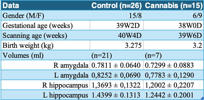
Comparison of amygdala and hippocampus volumes in control (black) and cannabis-exposed (gray) groups. Right amygdala volume is significantly lower in the cannabis group (*p=0.04), with no significant difference on the left (ns, p=0.2288). Right hippocampal volume shows a non-significant decrease (p=0.05), while the left hippocampus is significantly smaller in the cannabis group (*p=0.0113). Error bars represent the standard error of the mean (SEM).