
Environmental Health 1
Session: Environmental Health 1
 photo")
Vladislav Obsekov, MD (he/him/his)
Pediatric Cardiology Fellow
Mount Sinai
New York, New York, United States
.png) Parent phthalate and phthalate metabolites measured in maternal urine during pregnancy
Parent phthalate and phthalate metabolites measured in maternal urine during pregnancy Demographics
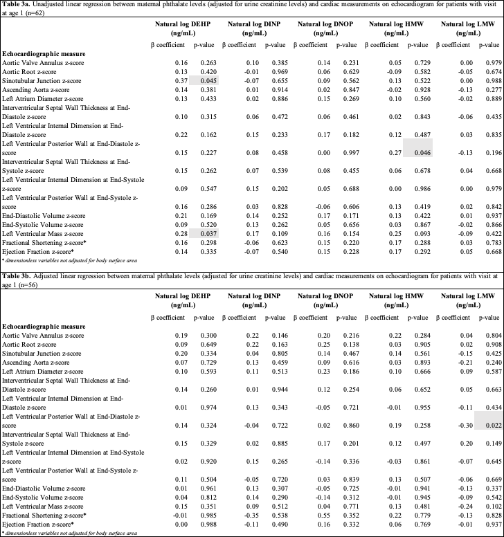 Linear regression