
Neonatal Neurology 5: Fetal
Session: Neonatal Neurology 5: Fetal
 photo")
Claire Baldauf, MD, MS (she/her/hers)
Assistant Professor
Children's Hospital Los Angeles
Los Angeles, California, United States
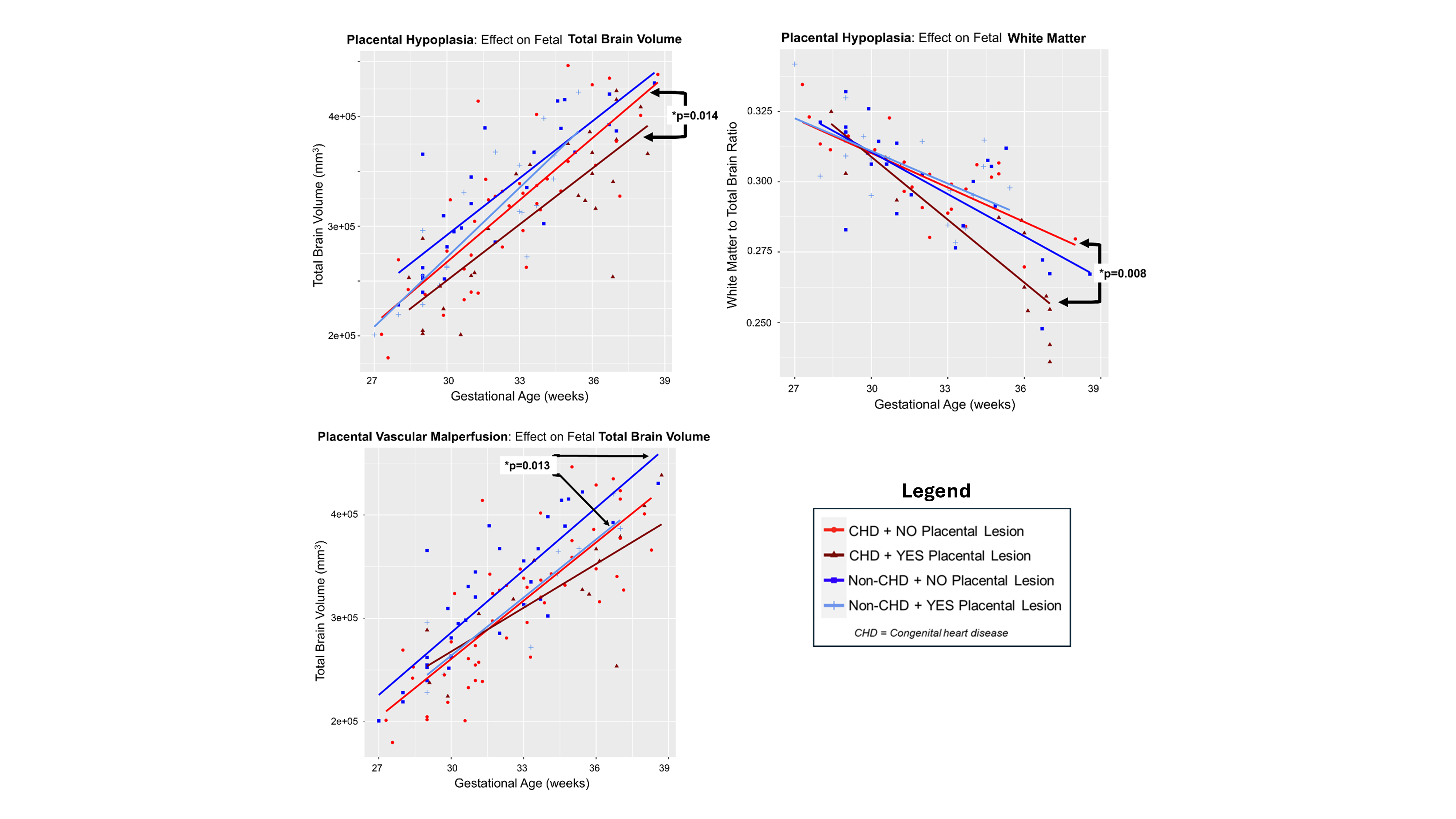 The effect of placental hypoplasia and placental vascular malperfusion on brain volume in fetuses with congenital heart disease (CHD) and those without (non-CHD). A, B) Within the CHD cohort, the presence of placental hypoplasia (defined as placental weight < 10th percentile) is associated with significantly lower total fetal brain volume (p=0.014) and reduced normalized fetal white matter volume (p=0.008). Non-CHD subjects did not show a significant difference between those with and without placental hypoplasia. C) Placental vascular malperfusion (either maternal, fetal, or both) was associated with lower total fetal brain volume in the combined cohort (inclusive of fetuses with and without CHD) (p=0.043), as well as the non-CHD cohort in stratified analysis (p=0.013). Fetuses affected by both CHD and placental vascular malperfusion demonstrated the smallest total brain volumes. The effect of placental hypoplasia and placental vascular malperfusion on brain volume in fetuses with congenital heart disease (CHD) and those without (non-CHD). A, B) Within the CHD cohort, the presence of placental hypoplasia (defined as placental weight < 10th percentile) is associated with significantly lower total fetal brain volume (p=0.014) and reduced normalized fetal white matter volume (p=0.008). Non-CHD subjects did not show a significant difference between those with and without placental hypoplasia. C) Placental vascular malperfusion (either maternal, fetal, or both) was associated with lower total fetal brain volume in the combined cohort (inclusive of fetuses with and without CHD) (p=0.043), as well as the non-CHD cohort in stratified analysis (p=0.013). Fetuses affected by both CHD and placental vascular malperfusion demonstrated the smallest total brain volumes.