677 - White Matter Microstructural Disruptions in Zika Virus-Exposed Children Without Congenital Zika Syndrome
Sunday, April 27, 2025
8:30am – 10:45am HST
Publication Number: 677.7092
Maria G.. Mora Alvarez, Children's National Hospital, Washington, DC, United States; Sarah B. Mulkey, Children's National Health System, Washington, DC, United States; Kushal Kapse, Children's National Health System, Washington, DC, United States; Yao Wu, Children's National Hospital, Washington, DC, United States; Hironori Teramoto, Children's National Health System, Arlington, VA, United States; Regan Andringa-Seed, Children's National Health System, Washington, DC, United States; Margarita Arroyave-Wessel, Children's National Health System, Washington, DC, United States; Madison Berl, Children's National Hospital, Washington, DC, United States; Raul E. Tarud Sabbag, Sabbag Radiologos, Barranquilla, Atlantico, Colombia; Gilbert Vezina, George Washington University School of Medicine and Health Sciences, Washington, DC, United States; Carlos A. Cure, BIOMELAB SAS, Barranquilla, Atlantico, Colombia; Josepheen De Asis-Cruz, Children's National Health System, Washington DC, DC, United States; Catherine Limperopoulos, Children's National Health System, Washington DC, DC, United States
Staff Scientist II Children's National Hospital Washington, District of Columbia, United States
Background: Preschool-aged children exposed to Zika virus (ZIKV) antenatally, but without microcephaly or congenital Zika syndrome (CZS), have differences in multiple domains of neurodevelopment, compared to non-exposed controls (NEC). Quantitative neuroimaging studies in children can investigate the neural mechanisms underlying these changes. Objective: To compare brain microstructural organization in major white matter (WM) tracts in normocephalic 7-year-old children with antenatal ZIKV exposure and NEC. Design/Methods: We analyzed whole-brain diffusion tensor imaging (DTI; 27 directions, b=1000 s/mm2) data from 52 7-year-old children, acquired in Colombia on a GE 1.5T MRI scanner and preprocessed with an optimized DTI pipeline. Diffusion metrics were measured in 11 WM tracts and their subdivisions, totaling 19 regions (Figure 1). Using analysis of covariance, with age and sex as covariates, we compared fractional anisotropy (FA), mean diffusivity (MD), radial diffusivity (RD), and axial diffusivity (AD) between NEC and ZIKV exposed children. A sub-analysis was also conducted to assess hemispheric differences. Bonferroni-Holm method was used to correct for multiple comparisons. Results: There was lower MD and AD in the Superior Longitudinal Fasciculus (SLF), Fornix, Anterior Thalamic Radiation (ATR), Cingulate Gyrus (CG), and Hippocampal Cingulum (HC), and lower RD in the SLF, Fornix, ATR, and CG in ZIKV-exposed children (n=37; 23 F) versus NEC (n=15; 9 females [F]). No significant differences were observed in FA values between the two groups. Brain laterality sub-analysis on whole sample revealed that the higher MD in ATR was only significant in the left hemisphere. The findings in the SLF and ATR survived multiple comparisons correction.
Conclusion(s): Although higher MD and RD values often reflect alterations in WM microstructure, this may not hold consistently in pediatric populations, where WM tracts are still undergoing significant development. The elevated MD, RD, and AD values observed in the SLF and ATR in NEC compared to ZIKV-exposed children may indicate active WM maturation in NEC. Future studies will correlate these diffusion measurements with neurodevelopmental assessments to enhance understanding of these findings. This study provides preliminary evidence of microstructural WM differences between ZIKV-exposed children without CZS and NEC.
Figure 1. Analyzed regions of interest (ROIs) in major white matter (WM) tracts. 
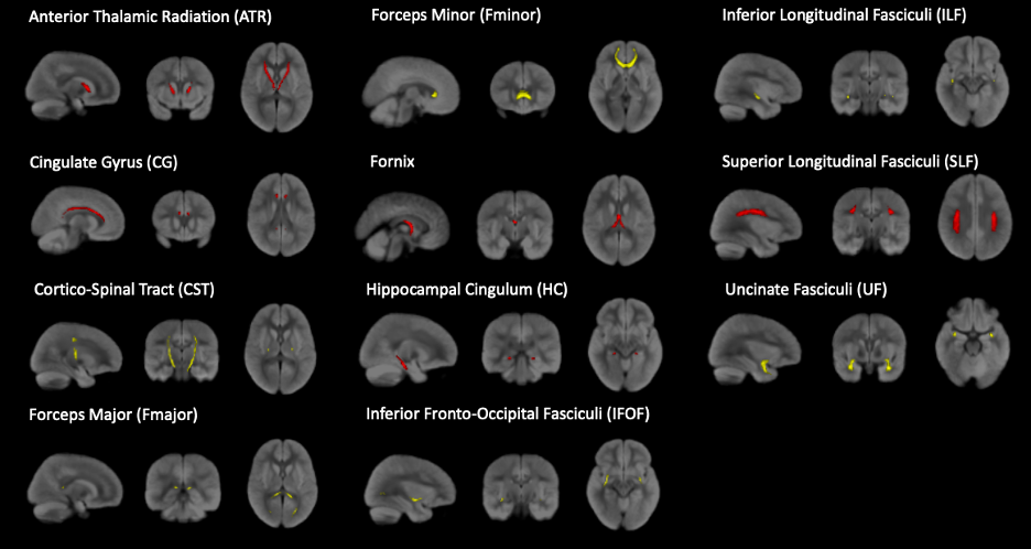
These regions were defined based on an age-specific probabilistic WM tract atlas for children aged 6-8 years. Tracts of interest were thresholded at p>0.6, except for the inferior longitudinal fasciculus, where a threshold of p>0.4 was applied. ROIs where DTI differences were detected between unexposed and antenatal zika virus-exposed children are shown in red.
Table 1. DTI differences between antenatal ZIKV exposed and NEC. 
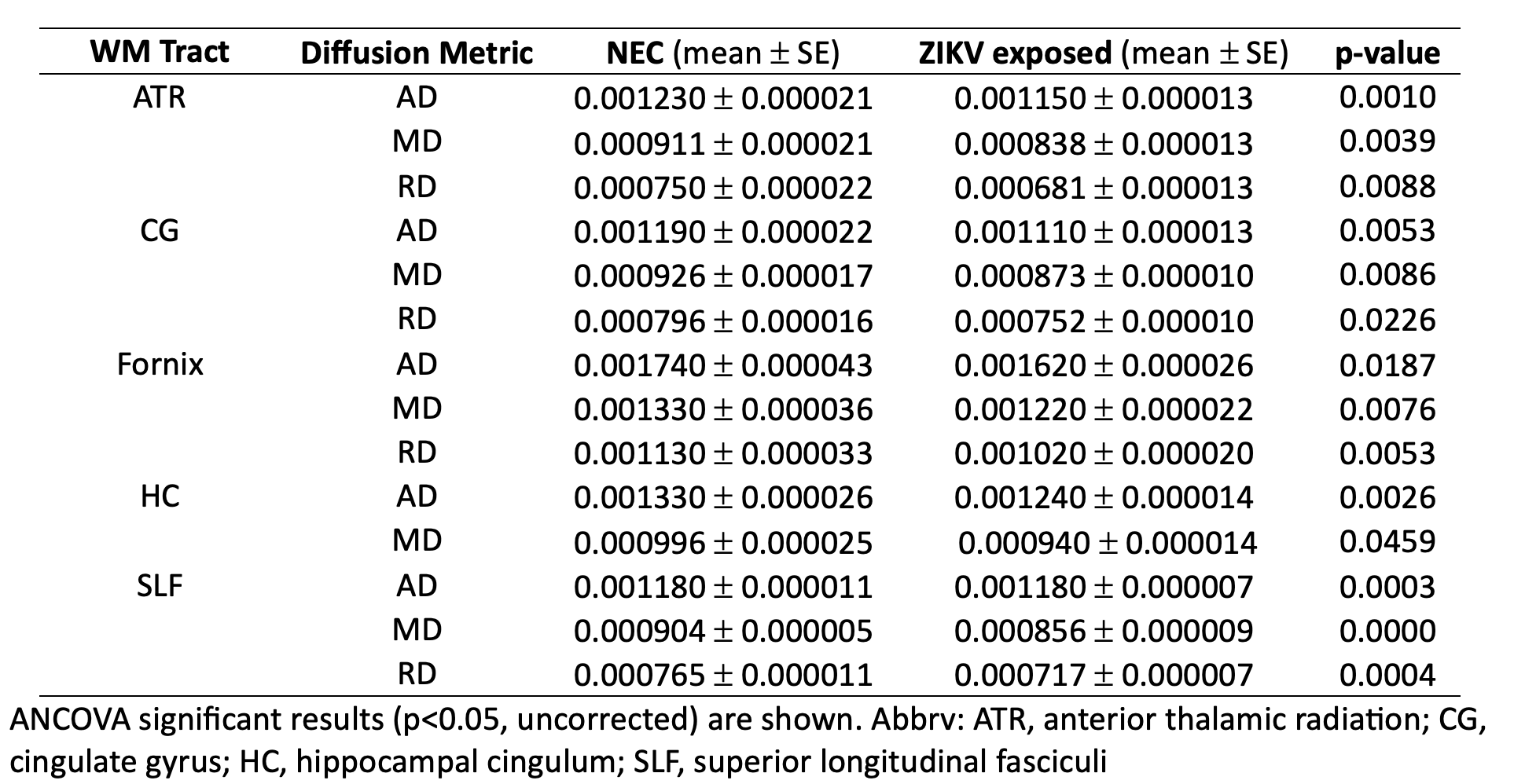
Covariates in analysis of covariance: sex and age. Adjusted means and significant p-values are displayed.