655 - Relationship Between Brain Temperature and Long-Term Outcomes of Infants with Hypoxic-Ischemic Encephalopathy
Sunday, April 27, 2025
8:30am – 10:45am HST
Publication Number: 655.6324
Natalie L. Smith, Children's Hospital Los Angeles, Los Angeles, CA, United States; Eni Jano, Children's Hospital Los Angeles, Los Angeles, CA, United States; Benita Tamrazi, CHLA, Los Angeles, CA, United States; Stefan Bluml, Children's Hospital Los Angeles/USC, Los Angeles, CA, United States; Tai-Wei Wu, University of Southern California, Keck School of Medicine, Los Angeles, CA, United States
Neonatal Perinatal Medicine Fellow University of Southern California Los Angeles, California, United States
Background: Infants with suspected hypoxic ischemic encephalopathy (HIE) undergoing whole-body hypothermia, have differences in regional brain temperatures, with the deeper structures having significantly lower temperatures. It is unknown if the temperatures differences are associated with long-term developmental outcomes. Objective: To determine the relationship between brain temperature during therapeutic hypothermia (TH) and long-term outcomes of infants with HIE. Design/Methods: This is a prospective observational study of Infants with suspected HIE who had MRI during TH. Regional brain temperature (basal ganglia-BG, thalamus-Thal, parietal white matter-WM, and cortical gray matter-cGM) was measured by MR thermometry during TH. MR injury severity (normal-mild or moderate-severe) and pattern (no injury, basal ganglia/thalamus-BGT, or watershed-WS) were scored by pediatric neuroradiologist using Barkovich scoring system. The primary outcome was death or neurodevelopmental impairment, which was defined as gross motor function classification system level of ≥1, diagnosis of cerebral palsy, or a cognitive score < 90 on the Bayley Scales of Infant and Toddler Development 3rd edition, at 12 to 36 months of age. This was designated as poor outcome. Statistics: Regional brain temperature were compared according to MR injury severity and pattern by unpaired t-test or Mann-Whitney test when non-normal. Logistic regression was used to assess relationship between brain temperature, MR injury severity, and outcome. Results: Overall, 55 patients (17 with poor outcomes) were included in the analysis, Table 1. The mean age when MRI was performed during TH was 2.3 ± 0.6 days. Rectal temperature was 33.3 ± 0.4ºC during MRI. There were no differences in regional brain temperature in patients with good versus poor outcome, except for a significantly higher Thal temperature in poor outcome group (33.9 vs. 33.0ºC, p< 0.01), Figure 1. Regional brain temperature of the WM and cGM were also significantly higher in those with WS compared to BG-T injury, Figure 2. No significant relationships between regional brain temperature and outcome were found based on the logistic regression model (controlling for injury severity, pattern, cord or first hour pH, and 5-minute Apgar).
Conclusion(s): In-vivo measurement of brain temperature reveal regional tissue resistance to cooling that is associated with injury pattern. However, brain temperature was not a predictor of long-term outcome.
Figure 2 
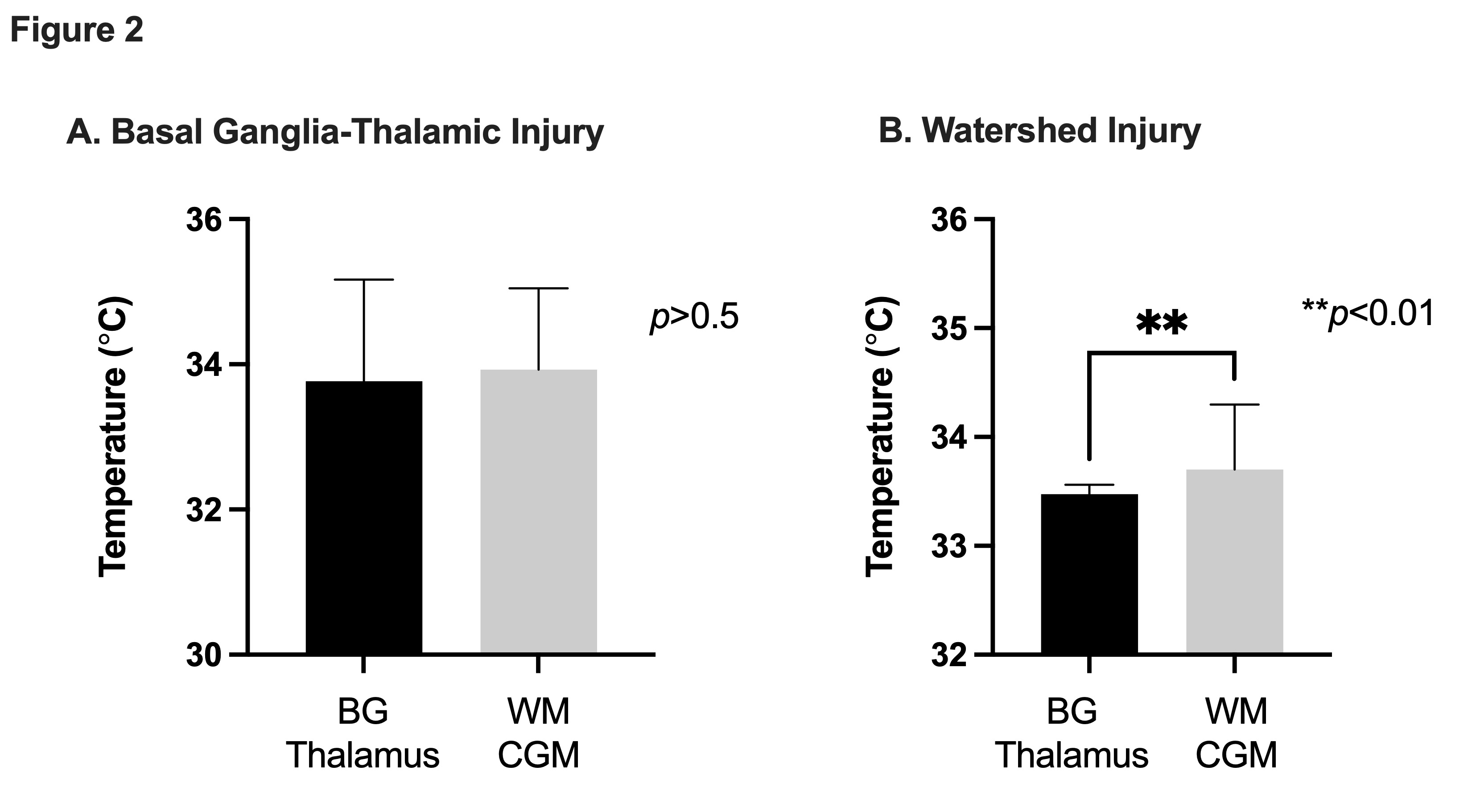
Regional brain temperatures compared to pattern of brain injury.
Table 1. Patient characteristics 
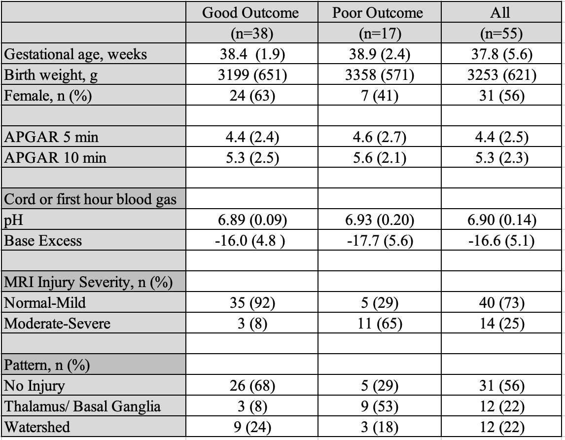
Numbers are reported as mean (standard deviation), unless specified as number (%).