
Obesity 2
Session: Obesity 2

Charles Herrin, MD
Physician
University of Texas Southwestern Medical Center
Dallas, Texas, United States
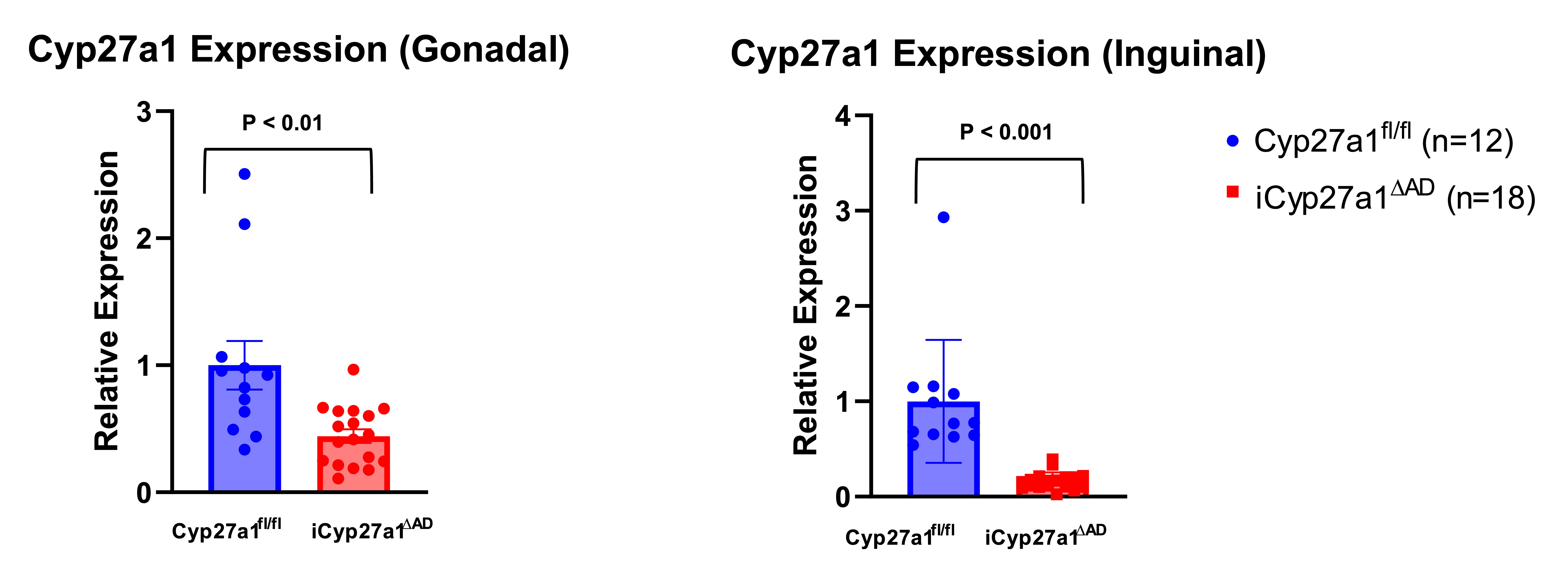 Changes in cellular Cyp27a1 expression in adipocytes. vWAT and sWAT adipocytes were analyzed by RT-qPCR. N=12,18 (p < 0.01 and p<0.001 respectively). Unpaired t-test.
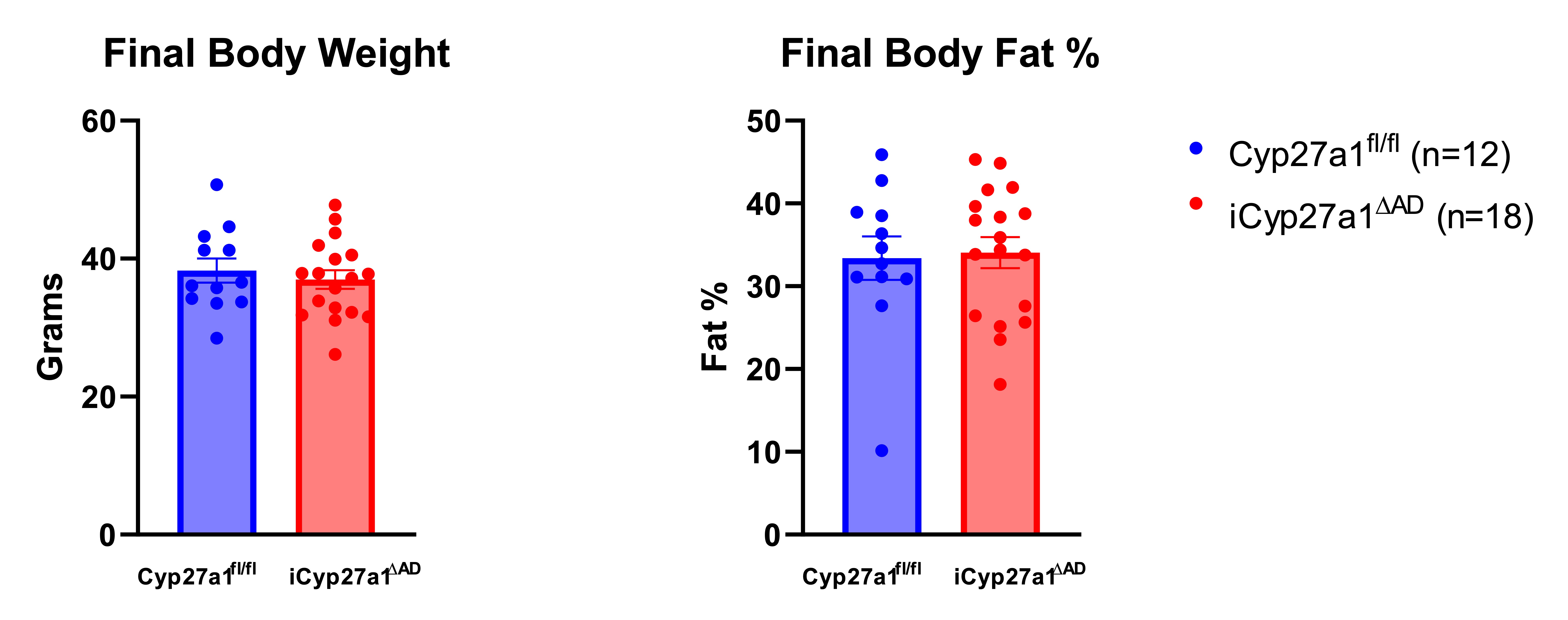 Body compositions of Cyp27a1fl/fl and iCyp27a1∆AD. Cyp27a1fl/fl and iCyp27a1∆AD were harvested after 12 weeks on HFD, final body weights were taken and final body fat was measured by NMR N=12,18 (p>0.99). Unpaired t-test.
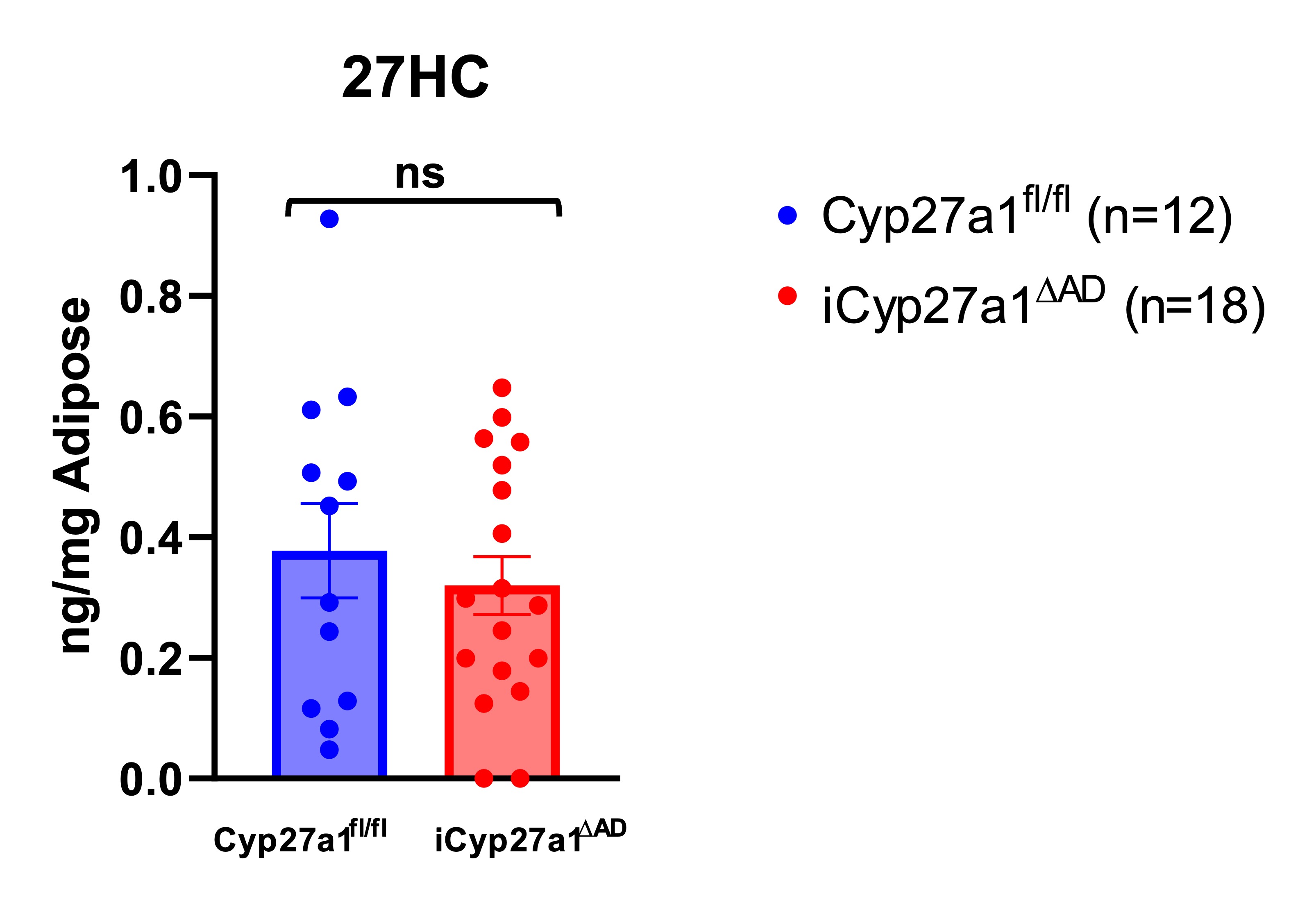 Cellular 27HC content in adipose tissue. Cyp27a1fl/fl and iCyp27a1∆AD were fed chow or HFD for 12 weeks, and vWAT were isolated for measurement of oxysterol concentrations using LC/MS. N=12,18. Unpaired t-test.Changes in cellular Cyp27a1 expression in adipocytes. vWAT and sWAT adipocytes were analyzed by RT-qPCR. N=12,18 (p < 0.01 and p<0.001 respectively). Unpaired t-test.Body compositions of Cyp27a1fl/fl and iCyp27a1∆AD. Cyp27a1fl/fl and iCyp27a1∆AD were harvested after 12 weeks on HFD, final body weights were taken and final body fat was measured by NMR N=12,18 (p>0.99). Unpaired t-test.Cellular 27HC content in adipose tissue. Cyp27a1fl/fl and iCyp27a1∆AD were fed chow or HFD for 12 weeks, and vWAT were isolated for measurement of oxysterol concentrations using LC/MS. N=12,18. Unpaired t-test.