Session: Neonatal General 9: Hematology, Bilirubin and Feeding
699 - Early Life Exposure to Hyperoxia Associated with Liver Injury in Newborn Rat Model
Saturday, April 26, 2025
2:30pm – 4:45pm HST
Publication Number: 699.4705
Mane Sargsyan, Cohen Children's Medical Center, New York, NY, United States; Fatima Toor, Cohen Children's Medical Center, New Hyde Park, NY, United States; Abigail Akselrod, Feinstein Institutes for Medical Research, East Meadow, NY, United States; Chunfang Zhao, Feinstein Institutes for Medical Research, New Hyde Park, NY, United States; Mansoor Nasim, New York University Long Island School of Medicine, Mineola, NY, United States; Shahana Perveen, Cohen Children Medical Center New york, Manhasset, NY, United States
Neonatology Fellow Cohen Children's Medical Center Glen Oaks, New York, United States
Background: Hyperoxia exposure is common in neonatal intensive care units for treating respiratory failure in preterm infants. While its pulmonary effects are well-known, the impact of prolonged high oxygen levels on the liver is not studied well. Objective: To explore the role of hyperoxia and liver injury during the early neonatal period in the rat model. Design/Methods: Newborn Sprague-Dawley rat pups were divided into two groups after delivery: a control group maintained in room air and a hyperoxia group exposed to 60% oxygen from birth until 14 days of life. All pups were exposed to the normal day light cycle. On day 14, the pups were euthanized. Liver tissues and blood were collected for histopathology and markers, respectively. Vascular endothelial growth factor (VEGF) and hepatocyte growth factor (HGF) were measured using quantitative real-time-PCR (RT-qPCR). Serum samples were analyzed for cytokines and liver injury markers. Results: Histopathological study revealed significant signs of liver cell injury and inflammation in the hyperoxia-exposed group compared to control group. Hyperoxic liver tissues showed increased hepatocyte mitosis, presence of eosinophils and multinucleated cells, perivenular inflammation, perisinusoidal vascular congestion, micro vesicular steatosis (an early sign of fatty liver changes), and increased apoptosis.
Quantitative real-time PCR showed that hepatocyte growth factor (HGF) gene expression was significantly elevated in the hyperoxia group (P < 0.01). The cytokine assay indicated a decrease in anti-inflammatory interleukin 10 (IL-10) levels (P < 0.001) and a reduction in interleukin 13 (IL-13), a modulator of hepatic injury (P < 0.05), in the hyperoxia-exposed pups. Vascular endothelial growth factor (VEGF) levels were markedly decreased in the hyperoxia group (P < 0.0001). Serum analysis demonstrated an increase in sorbitol dehydrogenase (SDH), an indicator of oxidative stress and liver cell damage, in the hyperoxia group compared to the control group.
Conclusion(s): Early life hyperoxia exposure in neonatal rat pups could result in significant hepatic injury, as evidenced by histopathological changes and markers of cellular damage and inflammation. This study paves the way for more targeted research for optimization of oxygen level in neonatal therapeutic strategies to mitigate potential risk and to lead to better clinical outcomes.
Multinucleated Cells 
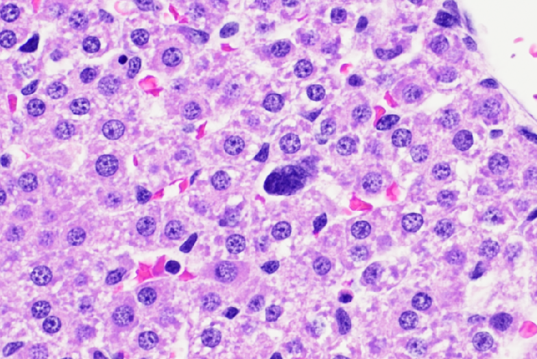
Haematoxylin-eosin staining of neonatal rat livers exposed to hyperoxia: presence of multinucleated cells
Mitosis and Eosinophils Haematoxylin-eosin staining of neonatal rat livers exposed to hyperoxia: increased mitosis in hepatocytes, presence of eosinophils.