729 - Redefining Bowel Wall Thickness Benchmarks for Preterm Infants
Saturday, April 26, 2025
2:30pm – 4:45pm HST
Publication Number: 729.6503
Maggie Vogel, Tufts Medical Center, Boston, MA, United States; INDRANI BHATTACHARJEE, Tufts University School of Medicine, Boston, MA, United States; Michael T. Dolinger, Icahn School of Medicine at Mount Sinai, New York, NY, United States
Neonatology Fellow Tufts Medical Center Boston, Massachusetts, United States
Background: Diagnosing gut pathology in premature infants is critical yet challenging, with early intervention essential for improved outcomes. Ultrasound is emerging as a promising tool for earlier detection of gastrointestinal abnormalities compared to radiographs. Necrotizing Enterocolitis (NEC) remains a severe threat, progressing rapidly from inflammation to ischemia. Current bowel wall thickness (BWT) standards (1-2.7 mm) are derived from pediatric data, lacking neonatal-specific validation. Neonatal BWT benchmarks could transform early diagnosis and management of critical GI pathologies in this vulnerable population. Objective: Using serial bowel ultrasound to longitudinally assess bowel wall thickness and inter-quadrant variation to create gestational age-based normative values. Design/Methods: An IRB-approved prospective cohort study was conducted at Tufts NICU in preterm infants under 34 weeks with an expected stay of more than two weeks. Weekly bedside abdominal ultrasounds were obtained from birth until eight weeks postnatal age or discharge, whichever came first. Abdominal quadrants were assessed systematically with a focus on identifying BWT in each of the four quadrants. All images were reviewed by a pediatric radiologist. Results: In this study, 14 infants, with a mean gestational age (GA) of 29.7 ± 2.9 weeks and a mean birth weight of 1371g ± 571g, as illustrated in Table 1 were included. BWT increased with GA across all quadrants, with no significant inter-quadrant variation (ANOVA p=0.102). Mixed- effects models confirmed GA as a significant BWT predictor (p < 0.001). Average BWT in preterm infants was 0.18-0.26 mm, much below normative pediatric values (Figure 1, 2).
Conclusion(s): This study presents gestational age-based BWT standards for preterm neonates facilitating precise GI health monitoring. The uniform BWT growth across quadrants supports a streamlined measurement approach underscoring the need for neonatal-specific standards. This study establishes neonatal-specific BWT benchmarks to aid early detection of bowel injury in preterm infants. Future research should focus on validating these BWT benchmarks across diverse neonatal populations and exploring the integration of ultrasound-derived BWT metrics with other clinical indicators of preterm bowel injury.
Table 1 Demographic and perinatal characteristics of study population. All data expressed in percentages unless otherwise specified. *p values significant at <0.05
Figure 1 
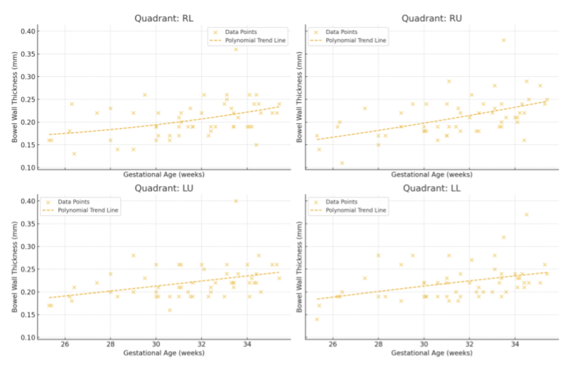
Scatter plots with trend lines for each abdominal quadrant showing bowel wall thickness by gestational age
Figure 2 
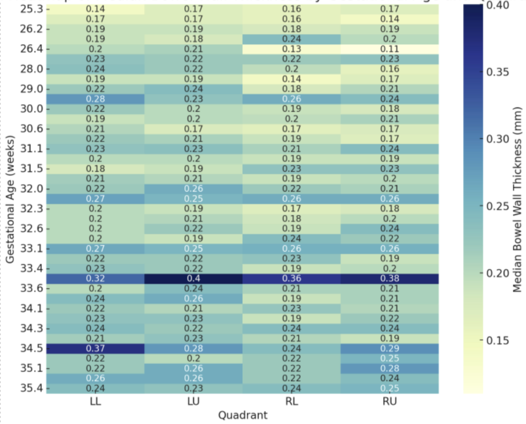
Heatmap of median bowel wall thickness by gestational age and quadrant