
Neonatal Hemodynamics and Cardiovascular Medicine 3
Session: Neonatal Hemodynamics and Cardiovascular Medicine 3
 photo")
Judith Leyens, Pediatrician, Neonatal-Perinatal Medicine Fellow (she/her/hers)
Neonatal-Perinatal Medicine Fellow
Department of Neonatology and Pediatric Intensive Care, University of Bonn, Germany; Division of Neonatology, Department of Pediatrics, BC Women´s and Children´s Hospital, University of British Columbia, Canada
Vancouver, British Columbia, Canada
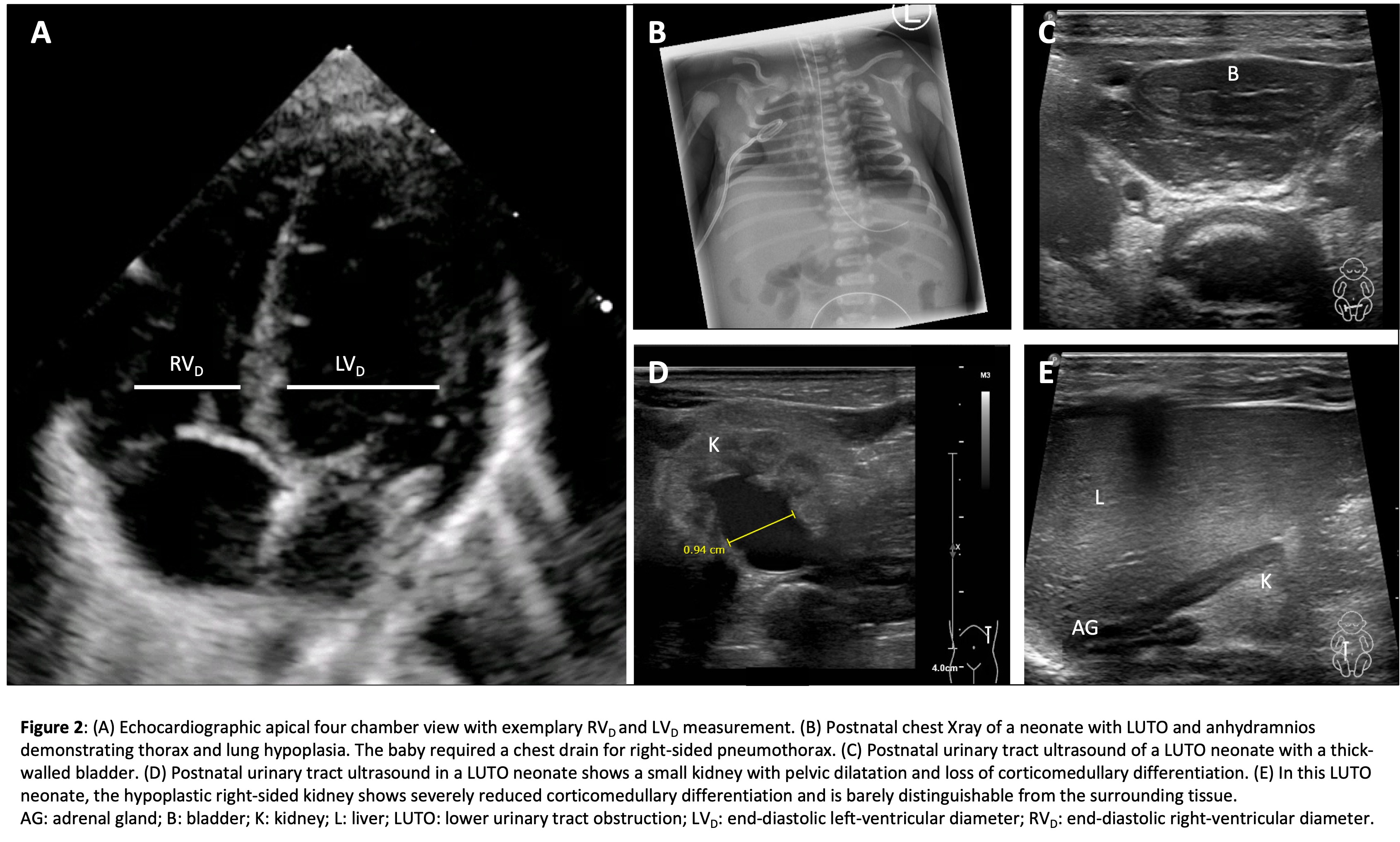 (A) Echocardiographic apical four chamber view with exemplary RVD and LVD measurement. (B) Postnatal chest Xray of a neonate with LUTO and anhydramnios demonstrating thorax and lung hypoplasia. The baby required a chest drain for right-sided pneumothorax. (C) Postnatal urinary tract ultrasound of a LUTO neonate with a thick-walled bladder. (D) Postnatal urinary tract ultrasound in a LUTO neonate shows a small kidney with pelvic dilatation and loss of corticomedullary differentiation. (E) In this LUTO neonate, the hypoplastic right-sided kidney shows severely reduced corticomedullary differentiation and is barely distinguishable from the surrounding tissue. _x000B_AG: adrenal gland; B: bladder; K: kidney; L: liver; LUTO: lower urinary tract obstruction; LVD: end-diastolic left-ventricular diameter; RVD: end-diastolic right-ventricular diameter.