043 - Exploring Structural Brain Metrics, Symmetry and Associations with Disease Features in Patients with Localized Craniofacial Scleroderma: A Structural Magnetic Resonance Imaging Study
Monday, April 28, 2025
7:00am – 9:15am HST
Publication Number: 43.5053
Diana Valdes Cabrera, The Hospital for Sick Children, Toronto, ON, Canada; Daniel H. Glaser, Yale School of Medicine, New Haven, CT, United States; Elena deRosas, University of Pittsburgh School of Medicine, Pittsburgh, PA, United States; Haley J. Havrilla, University of Pittsburgh School of Medicine, Pittsburgh, PA, United States; Asha Jeyanathan, The Hospital for Sick Children, Toronto, ON, Canada; Fernando Osorio Duque, UPMC Childrens Hospital of Pittsburgh, Pittsburgh, PA, United States; Subramanian Subramanian, UPMC Children’s Hospital of Pittsburgh, Pittsburgh, PA, United States; Kathryn Torok, UPMC Childrens Hospital of Pittsburgh, doylestown, PA, United States; Andrea Knight, The Hospital for Sick Children, Toronto, ON, Canada
Associate Professor of Pediatrics The Hospital for Sick Children Toronto, Ontario, Canada
Background: Craniofacial localized scleroderma (Cf-LS) is a rare autoimmune disorder that affects face and scalp tissues and is commonly diagnosed during childhood. Children with Cf-LS may have brain involvement, often ipsilateral to facial lesions, which can be evaluated with magnetic resonance imaging (MRI). Existing neuroimaging studies are mainly limited to qualitative lesion assessments. Little is known about the extent of brain structure changes and association with other disease features. Objective: To examine interhemispheric brain volumetric differences regarding facial lesion sidedness in patients with Cf-LS utilizing structural MRI, and to evaluate if volumes associate with disease characteristics and patient quality of life. Design/Methods: Clinical T1w-MRI were extracted from patients with Cf-LS scanned at 1.5/3T at UPMC Children’s Hospital of Pittsburgh from 2003 to 2024. Brain metrics were automatically segmented and included left (L) / right (R) subcortical grey and white matter (GM, WM), cortical lobe, and cerebellar volumes. Facial sidedness was noted; then, asymmetry indices (L-R/(L+R)/2)) were calculated and interhemispheric differences were evaluated with paired t-tests. Associations between volumes and asymmetry indices, disease duration, activity, damage and severity (physician global assessment), and overall patient quality of life (visual analog scale), were evaluated with regression analyses adjusted for age, sex, total intracranial volume, and magnetic field. Results: Participants included 25 patients aged 5-21 years (Table 1). Interhemispheric differences were observed in the occipital lobe with 22 (88%) patients displaying lower volumes in the left hemisphere, irrespective of facial sidedness (T=-3.78, p< 0.001; Fig. 1a). Lower bilateral WM volumes (left: T=-2.39, p=0.027; right: T=-2.34, p=0.030) were associated with left facial sidedness (Fig 1b). Lower right occipital volumes were linked to greater disease damage (T=-2.79, p=0.014) and severity (T=-2.62, p=0.019). Higher left versus right subcortical GM volumes, given as a higher asymmetry index, were linked to better quality of life (T=-2.25, p=0.042).
Conclusion(s): In this cohort of youth with Cf-LS, we found interhemispheric differences and lower regional brain volumes associated with disease features, suggesting broad effects of Cf-LS on the brain. Larger studies, inclusive of controls, are needed to further characterize brain involvement in patients with Cf-LS and examine the role of structural brain MRI in monitoring disease progression.
Table 1: Patient Demographic and Disease Characteristics
Figure 1 
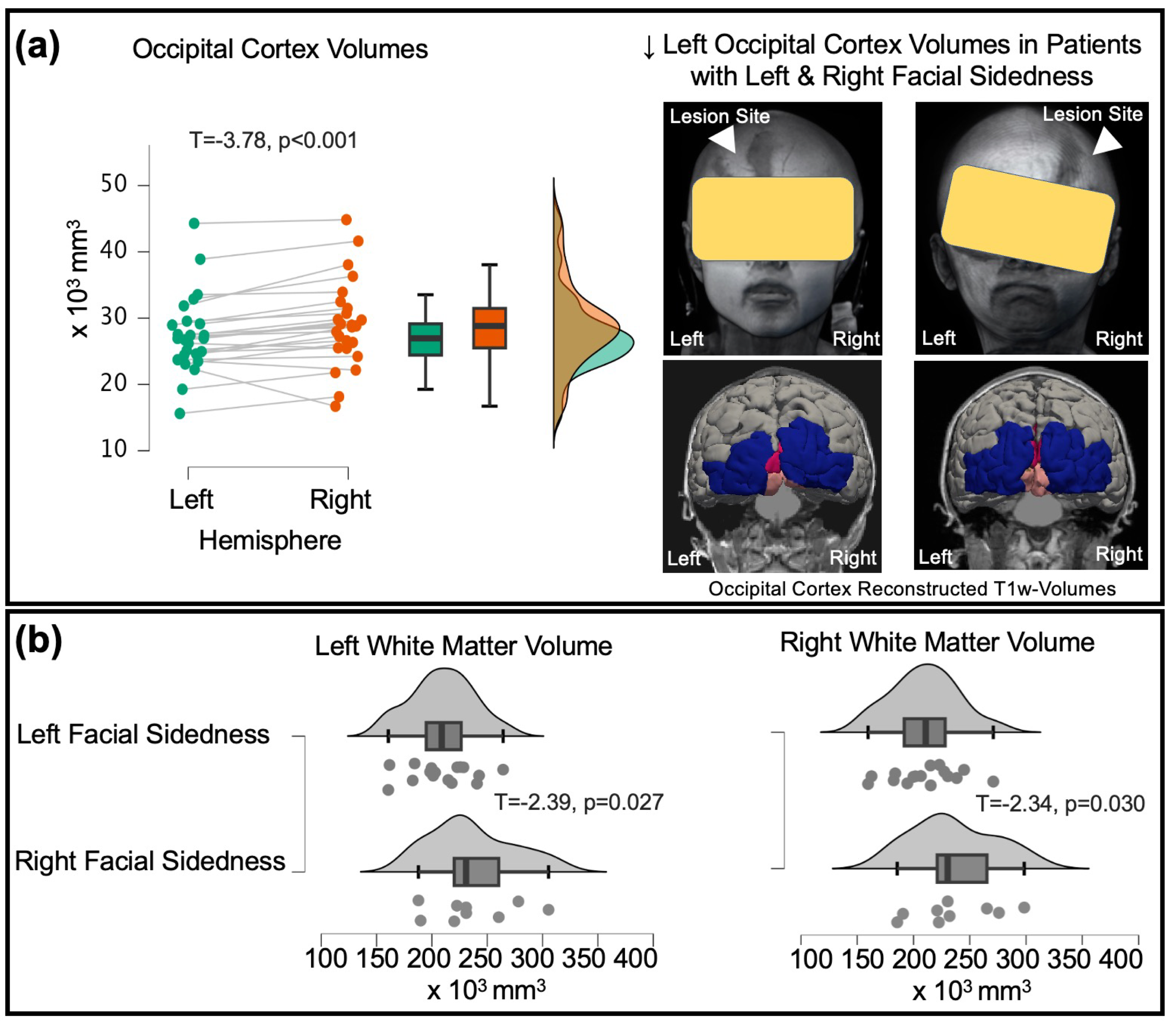
Figure 1: In patients with Cf-LS (a) lower occipital cortex volumes were observed in the left compared to the right hemisphere irrespective of facial lesion sidedness (coronal brain view of a 5-year-old female patient with face/scalp lesion in the left side and a 11-year-old female patient with face/scalp lesion in the right side displayed lower occipital volumes in left versus right hemisphere); while (b) lower left and right white matter volumes were associated with left facial sidedness.