
Neonatal Neurology 7: Pre-Clinical 1
Session: Neonatal Neurology 7: Pre-Clinical 1
Angela Saadat, PhD (she/her/hers)
Community Assistant Professor
Old Dominion University
NORFOLK, Virginia, United States
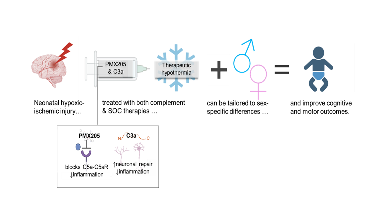 We hypothesized temporarily attenuating complement following hypoxic-ischemic injury in may lead to less neurological injury and improve functional outcomes in HIE. Further, we tested complement modulation with and without the SOC, TH, to determine if the two therapies synergize.
 .png) 3 days post-injury. a. Representative brain images and b. lesion area measurements demonstrate treatment with CT and TH each yielded smaller brain lesions compared to untreated rats, and combining the treatments further reduced lesion sizes. When the data were stratified by sex, females benefited from TH and not CT therapies, where males benefitted from CT but not TH, but both benefited from combination therapy. Total lesion area was calculated from hemispheric loss and white infarct area measurements. Floating bars represent median values, shaded bars represent absolute deviation. c. & d. Immunofluorescence demonstrates levels of inflammatory markers in coronal brain sections. Red bars indicate median (c) or mean (d) fluorescence with absolute deviation or standard error bars. * Signifies p-values <0.05>0.005, **p-value <0.005 by Steel-Dwass, Dunnett’s or Wilcoxan methods.
3 days post-injury. a. Representative brain images and b. lesion area measurements demonstrate treatment with CT and TH each yielded smaller brain lesions compared to untreated rats, and combining the treatments further reduced lesion sizes. When the data were stratified by sex, females benefited from TH and not CT therapies, where males benefitted from CT but not TH, but both benefited from combination therapy. Total lesion area was calculated from hemispheric loss and white infarct area measurements. Floating bars represent median values, shaded bars represent absolute deviation. c. & d. Immunofluorescence demonstrates levels of inflammatory markers in coronal brain sections. Red bars indicate median (c) or mean (d) fluorescence with absolute deviation or standard error bars. * Signifies p-values <0.05>0.005, **p-value <0.005 by Steel-Dwass, Dunnett’s or Wilcoxan methods.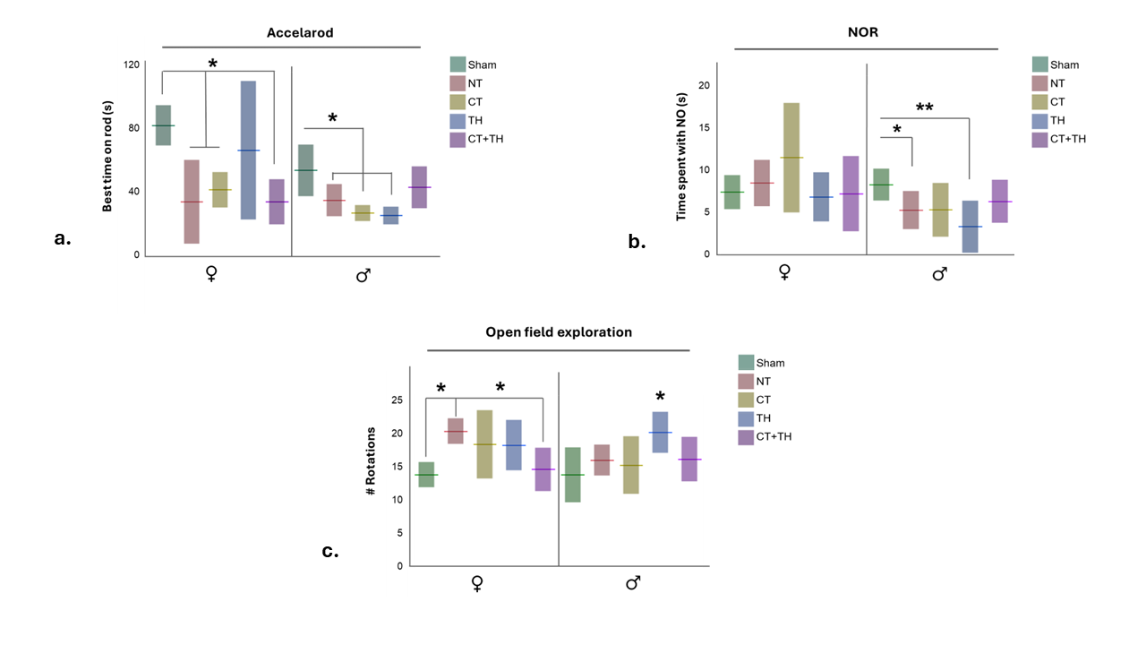 a. Rats were challenged to maintain locomotion on a rotating rod that accelerated in speed over 60s. b. Memory was tested in rats with the novel object recognition was test. c. General movement and exploration was assessed in the open field test. Floating bars indicate median (a) or mean (b & c), and shaded bars absolute deviation or standard deviation. * Signifies p-values <0.05>0.005, **p-value <0.005 by Wilcoxan or Tukey’s method.We hypothesized temporarily attenuating complement following hypoxic-ischemic injury in may lead to less neurological injury and improve functional outcomes in HIE. Further, we tested complement modulation with and without the SOC, TH, to determine if the two therapies synergize. 3 days post-injury. a. Representative brain images and b. lesion area measurements demonstrate treatment with CT and TH each yielded smaller brain lesions compared to untreated rats, and combining the treatments further reduced lesion sizes. When the data were stratified by sex, females benefited from TH and not CT therapies, where males benefitted from CT but not TH, but both benefited from combination therapy. Total lesion area was calculated from hemispheric loss and white infarct area measurements. Floating bars represent median values, shaded bars represent absolute deviation. c. & d. Immunofluorescence demonstrates levels of inflammatory markers in coronal brain sections. Red bars indicate median (c) or mean (d) fluorescence with absolute deviation or standard error bars. * Signifies p-values <0.05>0.005, **p-value <0.005 by Steel-Dwass, Dunnett’s or Wilcoxan methods.a. Rats were challenged to maintain locomotion on a rotating rod that accelerated in speed over 60s. b. Memory was tested in rats with the novel object recognition was test. c. General movement and exploration was assessed in the open field test. Floating bars indicate median (a) or mean (b & c), and shaded bars absolute deviation or standard deviation. * Signifies p-values <0.05>0.005, **p-value <0.005 by Wilcoxan or Tukey’s method.