
Neonatal Neurology 7: Pre-Clinical 1
Session: Neonatal Neurology 7: Pre-Clinical 1
 photo")
Michelle A. Sunshine, MD (she/her/hers)
Neonatology Fellow
Maria Fareri Children's Hospital at Westchester Medical Center
Dobbs Ferry, New York, United States
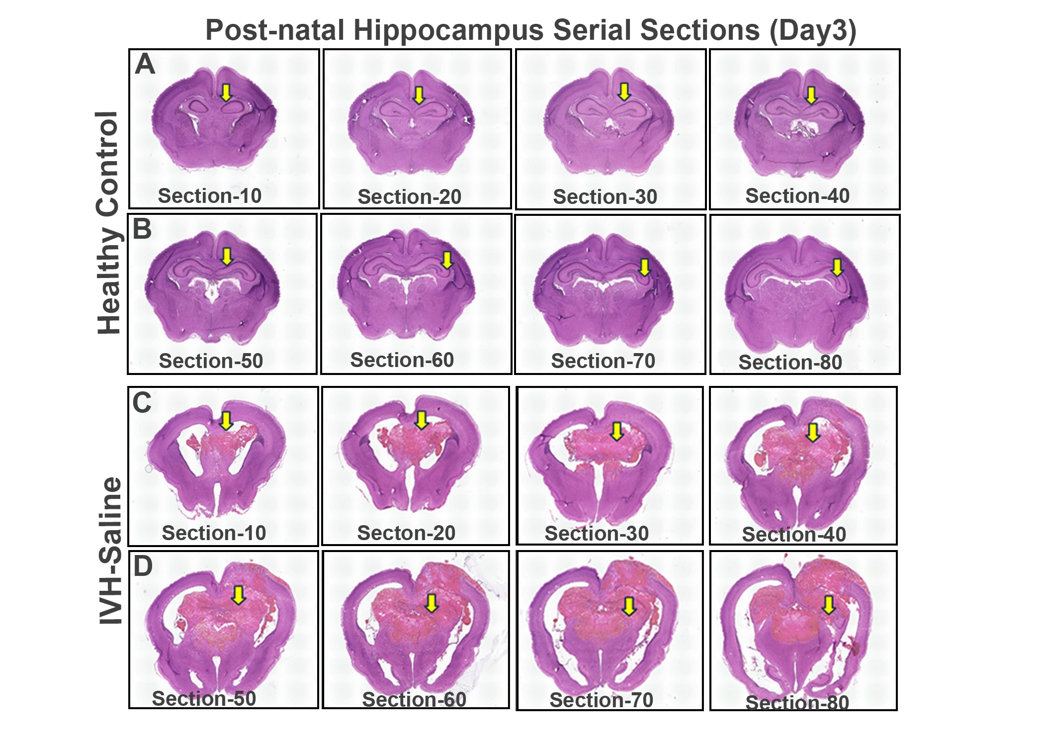 Hippocampal serial sections anterior to posterior for control vs vehicle treated + IVH groups showing parenchymal blood after IVH, tissue distortion and enlarged ventricles with arrows comparing similar regions.
 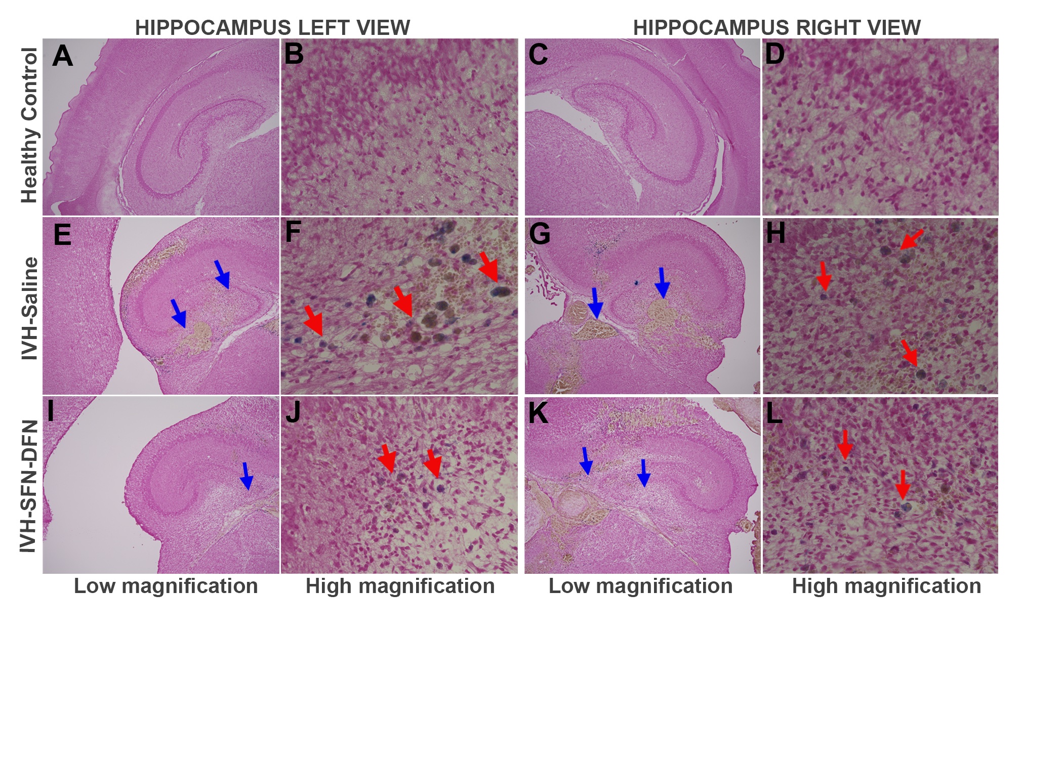 Iron deposition (arrows) in the hippocampus subregions for healthy control vs vehicle treated + IVH vs IVH + SFN-DFN treatment groups showing fewer positive cells (red arrows) after treatment (4x & 20x).Hippocampal serial sections anterior to posterior for control vs vehicle treated + IVH groups showing parenchymal blood after IVH, tissue distortion and enlarged ventricles with arrows comparing similar regions. Iron deposition (arrows) in the hippocampus subregions for healthy control vs vehicle treated + IVH vs IVH + SFN-DFN treatment groups showing fewer positive cells (red arrows) after treatment (4x & 20x).