143 - Tensor-Based Morphometry Identifies Impaired Growth of Novel Brain Areas in Fetuses with Congenital Heart Disease
Friday, April 25, 2025
5:30pm – 7:45pm HST
Publication Number: 143.6826
Yao Wu, Children's National Hospital, Washington, DC, United States; Josepheen De Asis-Cruz, Children's National Health System, Washington DC, DC, United States; Nickie Andescavage, Children's National Health System, Washington, DC, United States; Catherine Limperopoulos, Children's National Health System, Washington DC, DC, United States; Srikanth Damera, Children's National Health System, Mount Rainier, MD, United States
Medical Resident Children's National Hospital Mount Rainier, Maryland, United States
Background: The brains of fetuses with congenital heart disease (CHD) show morphological differences in the latter half of gestation which may predict their risk of future neurodevelopmental disability. However, studies showing this effect used manually labeled brain regions or studied macroscale differences at the level of large brain volumes, tissue classes, or lobes which may mask brain areas that are vulnerable in CHD. Tensor-based morphometry (TBM) allows the unbiased exploration morphological differences in fetuses with CHD versus controls. Objective: To compare morphological differences between the brains of fetuses with CHD and healthy controls using a novel TBM approach. Design/Methods: We analyzed 62 fetal T2 anatomical MRIs acquired between 28-30 weeks GA (23 CHD and 39 controls; Tbl. 1 ). Skull-stripped fetal brains were registered to a 29-week gestational age template using an affine transformation followed by a non-linear warp. TBM uses the log-determinant of the Jacobian maps generated by the non-linear warp to quantify changes in local brain volume. By comparing these maps between healthy controls fetuses and those with CHD while controlling for gestational age at scan and sex we can test, without any a priori assumptions, how brain volumes differ between the two groups. Results are voxel-wise corrected at the FDR 0.05 level with a minimum cluster-volume of 20mm3. Results: Using TBM, we show that CHD reduces fetal cerebellar, bilateral primary somatomotor, bilateral precuneus, bilateral orbitofrontal cortex, left medial prefrontal cortex, left insula, and right superior frontal cortex volumes. In contrast, the left superior parietal lobe, right lateral ventricle, between the horn and the trigone, and a portion of the right inferior temporal lobe were expanded in the brains of fetuses with CHD compared to those of controls.
Conclusion(s): Using TBM we identified areas of reduced fetal brain volume in CHD in a spatially unbiased manner – a key limitation of prior approaches. For instance, some previous studies may have found no effect of CHD in the cerebellum because analyzing the whole structure masked the localized findings identified here. Our results show that CHD causes dysmaturity in fetal brain regions essential for motor and cognitive functions which may lead to the common deficits in executive function and adaptive motor planning seen in children with CHD.
Table 1: Clinical Characteristics of the Cohort
Changes in Fetal Brain Volumes Due to CHD. 
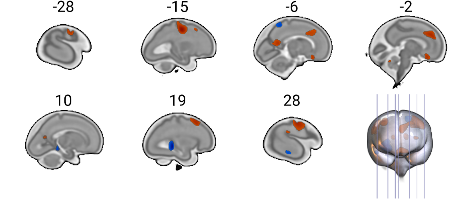
Brain areas in red indicate those that are larger in healthy control fetal brain compared to those with CHD. Conversely those in blue indicate those that are larger in CHD fetal brain compared to those of healthy controls. Results are voxel-wise FDR corrected.