
Cardiology 2
Session: Cardiology 2
 photo")
Christina M. Harris, M.S. (she/her/hers)
Student
Howard University College of Medicine
Hyattsville, Maryland, United States
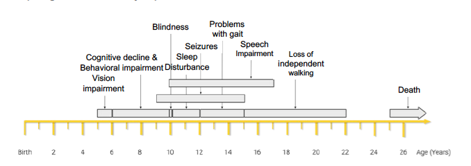 Each gray bar represents average age range of onset. [T.-R. (n.d.). Learn about batten disease & support from BDSRA Australia. BDSRA Australia.]
.png) Gray shaded areas depict the normal range of heart rates for the age intervals 0-1, 1-5, 5-10, 10-15, and > 15 years
Gray shaded areas depict the normal range of heart rates for the age intervals 0-1, 1-5, 5-10, 10-15, and > 15 years.png) Data point for the 80-year-old study participant was excluded for visualization purpose. The outlier increased the slope of the trendline. Gray shaded areas depict the normal range of heart rates for the age intervals 0-1, 1-5, 5-10, 10-15, and > 15 yearsEach gray bar represents average age range of onset. [T.-R. (n.d.). Learn about batten disease & support from BDSRA Australia. BDSRA Australia.]Gray shaded areas depict the normal range of heart rates for the age intervals 0-1, 1-5, 5-10, 10-15, and > 15 yearsData point for the 80-year-old study participant was excluded for visualization purpose. The outlier increased the slope of the trendline. Gray shaded areas depict the normal range of heart rates for the age intervals 0-1, 1-5, 5-10, 10-15, and > 15 years
Data point for the 80-year-old study participant was excluded for visualization purpose. The outlier increased the slope of the trendline. Gray shaded areas depict the normal range of heart rates for the age intervals 0-1, 1-5, 5-10, 10-15, and > 15 yearsEach gray bar represents average age range of onset. [T.-R. (n.d.). Learn about batten disease & support from BDSRA Australia. BDSRA Australia.]Gray shaded areas depict the normal range of heart rates for the age intervals 0-1, 1-5, 5-10, 10-15, and > 15 yearsData point for the 80-year-old study participant was excluded for visualization purpose. The outlier increased the slope of the trendline. Gray shaded areas depict the normal range of heart rates for the age intervals 0-1, 1-5, 5-10, 10-15, and > 15 years