403 - Nutrient Restriction Stimulates the Release of Anti-angiogenic microRNAs from Trophoblast Stem Cells in an In Vitro Fetal Growth Restriction Model
Friday, April 25, 2025
5:30pm – 7:45pm HST
Publication Number: 403.5747
Caitlin Eason, University of Colorado School of Medicine, Denver, CO, United States; JAMES R. BARDILL, UNIVERSITY OF COLORADO-ANSCHUTZ MED, AURORA, CO, United States; Anis karimpour-fard, University of Colorado School of Medicine, Aurora, CO, United States; Courtney Breckenfelder, CU Denver Anschutz Medical Campus, Aurora, CO, United States; Holly Wood, University of Colorado AMC, Thornton, CO, United States; Theresa L. Powell, University of Colorado, Denver, CO, United States; Clyde Wright, University of Colorado School of Medicine, Aurora, CO, United States; S. Christopher Derderian, University of Colorado School of Medicine, Denver, CO, United States; Madison Crew, Rocky Vista University School of Osteopathic Medicine, Parker, CO, United States
Pediatric Surgery Research Fellow University of Colorado School of Medicine Denver, Colorado, United States
Background: A hallmark pathological feature of fetal growth restriction (FGR) is reduced villous vascular branching in the placenta, which impairs nutrient and oxygen delivery to the fetus. While several animal and culture models exist to study FGR, those involving nutrient restriction reflect the phenotypic characteristics observed in human FGR. Human trophoblast stem cells (hTSCs), which are critical for early placental development, are impacted by nutrient availability, with restricted nutrient conditions potentially affecting their function and signaling. Epigenetic regulators, specifically microRNAs (miRNAs), are believed to contribute to this pathogenesis by modulating gene expression post-transcriptionally. These miRNAs can be transported through extracellular vesicles (EVs), allowing hTSCs to influence nearby or distant cells, including endothelial cells, which are essential for placental vascular development. We hypothesize that nutrient restricted hTSCs release miRNAs via EVs, targeting endothelial cells and downregulating angiogenic pathways, thus contributing to the vascular deficiencies characteristic of FGR placentas. Objective: Determine whether hTSCs exposed to nutrient restricted culture media upregulate expression of anti-angiogenic miRNAs released via EVs. Design/Methods: hTSCs were plated in either nutrient complete or a 50% nutrient restricted media. Forty-eight hours after nutrient restriction, we performed an MTT assay and measured CASP3 gene expression to determine if our model reduced proliferation without increasing apoptosis. Next, we isolated EVs from the two conditions and sequenced their miRNA cargo. miRNAs with significantly different expressions between nutrient-complete and -restricted were mapped using Ingenuity Pathway Analysis (IPA) to identify potential targets of angiogenic molecular pathways. Results: hTSCs exposed to a 50% nutrient restricted media had reduced hTSC proliferation without increased apoptosis, mimicking the metabolic stress associated with FGR (Figure 1). EV miRNA-seq identified three upregulated (miR-92a-3p, miR-142-3p, and miR-141-3p) and three downregulated (miR-517a-3p, miR-517b-3p, and miR-126-3p) miRNAs with significant differential expression (Figure 2A-B). IPA predicted that miR-141-3p and miR-142-3p inhibit multiple genes involved in angiogenic pathways (Figure 2C).
Conclusion(s): In a hTSC In Vitro model, nutrient-restriction results in the upregulation of anti-angiogenic miRNAs released from hTSC via EVs. We propose this is a mechanism for reduced angiogenesis in FGR. Future work will focus on endothelial targets of miR-141-3p and miR-142-3p.
Cellular Effects of Nutrient Restriction Human trophoblast stem cells (hTSCs) from three unique cell lines were exposed to nutrient complete media or media with a 50% reduction of nutrients. MTT (colorimetric assay for measuring cell metabolic activity and proliferation); CASP3, Caspase 3 (protein coding gene that plays a critical role in apoptosis); qPCR, quantitative polymerase chain reaction.
Extracellular Vesicle microRNA Sequencing 
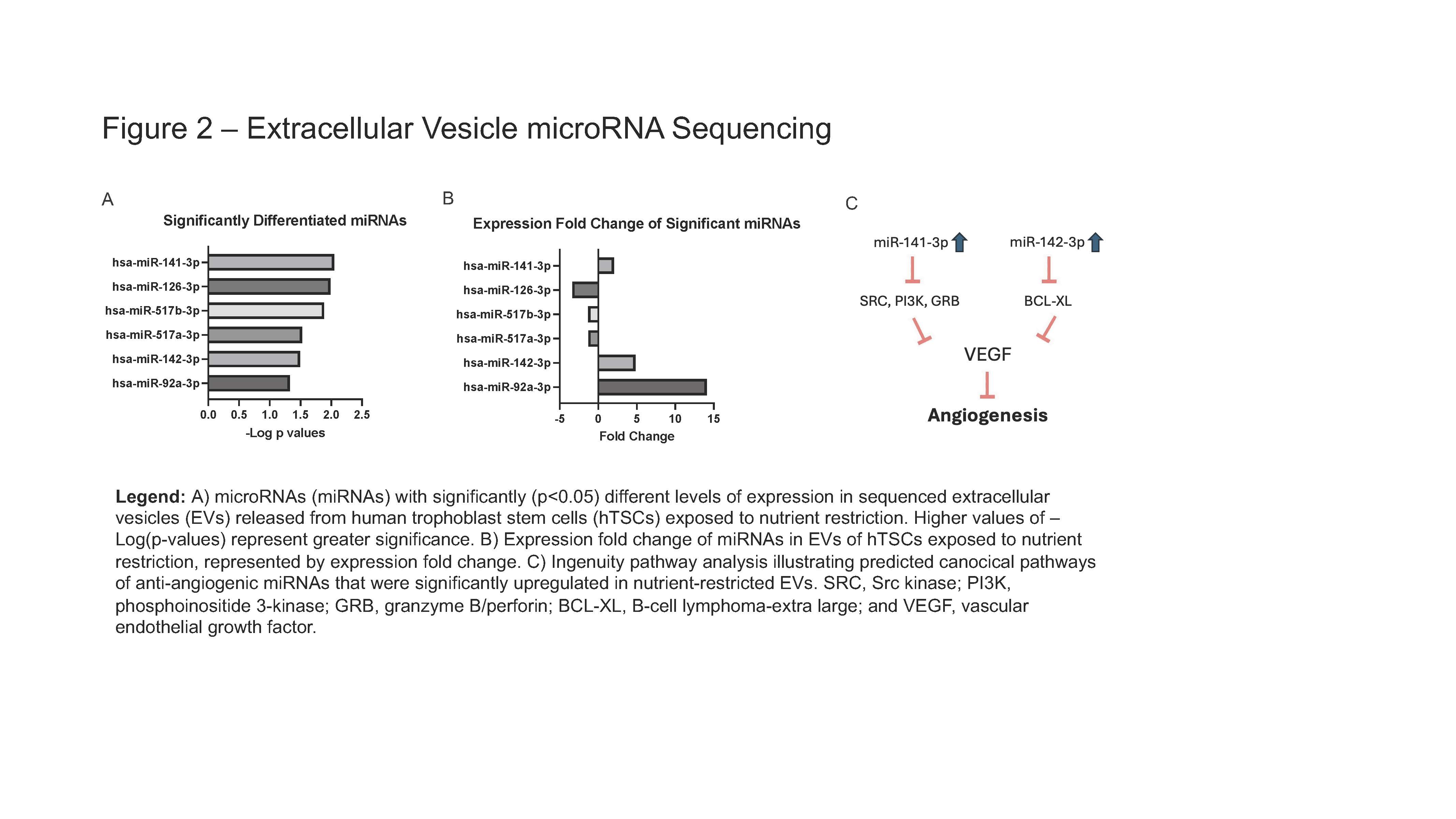
A) microRNAs (miRNAs) with significantly (p < 0.05) different levels of expression in sequenced extracellular vesicles (EVs) released from human trophoblast stem cells (hTSCs) exposed to nutrient restriction. Higher values of –Log(p-values) represent greater significance. B) Expression fold change of miRNAs in EVs of hTSCs exposed to nutrient restriction, represented by expression fold change. C) Ingenuity pathway analysis illustrating predicted canocical pathways of anti-angiogenic miRNAs that were significantly upregulated in nutrient-restricted EVs. SRC, Src kinase; PI3K, phosphoinositide 3-kinase; GRB, granzyme B/perforin; BCL-XL, B-cell lymphoma-extra large; and VEGF, vascular endothelial growth factor.