355 - The relationship between whole brain neural network development and cardiac physiology among infants born with critical congenital heart disease
Friday, April 25, 2025
5:30pm – 7:45pm HST
Publication Number: 355.6854
Lauren Harasymiw, University of California San Francisco, San Francisco, CA, United States; Amy Kuang, University of California, San Francisco, School of Medicine, Daly City, CA, United States; Megan Martin, Stanford University, Stanford, CA, United States; Duan Xu, UCSF, San Francisco, CA, United States; Elizabeth George, UCSF Benioff Children's Hospital San Francisco, San Francisco, CA, United States; Shabnam Peyvandi, UCSF Benioff Children's Hospital San Francisco, San Francisco, CA, United States; Patrick McQuillen, University of California, San Francisco, School of Medicine, San Francisco, CA, United States
Clinical Fellow University of California San Francisco San Francisco, California, United States
Background: Neonatal brain injury and brain dysmaturation are common among infants born with critical congenital heart disease (CCHD). Infants born with single ventricle physiology (SVP) may be at particularly high risk of abnormal brain development due to the palliative, staged surgical approach with prolonged cyanosis into childhood as compared to those with d-Transposition of the Great Arteries (d-TGA) who receive a definitive neonatal surgical repair. Global brain growth has been shown to proceed more slowly in newborns with SVP as compared to TGA. Objective: In this study, we used diffusion tensor imaging (DTI), in combination with graph theory to model neural connectivity, to evaluate the trajectory of neural network development across the newborn perioperative period in relationship to cardiac diagnosis. Design/Methods: Newborns with SVP and d-TGA were enrolled in a longitudinal cohort study and imaged pre- and post-operatively using DTI (3T MRI, 30 directions, b-value 700 s/mm2) in the neonatal period. Deterministic tractography was performed using Diffusion Toolkit, and ninety cerebral regions of interest were identified by mapping the Neonatal UNC Infant012 Atlas to 1 mm isotropic T1-SPGR images. Co-registration of the DTI and T1 images was performed using the FMRIB Linear Image Registration Tool (FLIRT). We constructed undirected adjacency matrices weighted by fractional anisotropy for each time point and then calculated graph theory metrics (global efficiency, modularity, transitivity, small-worldness) to quantify whole-brain network development. Mixed-effects linear models were used to compare the trajectory of brain development between cardiac lesion groups and those with and without brain injury. Results: 36 subjects were included (d-TGA = 16; SVP= 20). The sample was majority male (75%), and approximately a third (33.3%) of infants had significant white matter injury or stroke identified on their pre-operative MRI (Table 1). Neural network development did not differ significantly by brain injury status. In infants with SVP, the rate of global efficiency development was lower than those with d-TGA (t = -2.03, p = 0.044) after adjusting for age at scan (Figure 1). There were no differences in modularity, transitivity, or small-worldness across the cardiac lesion groups.
Conclusion(s): Infants with SVP had a slower increase in global efficiency during the newborn perioperative period, which may be related to the persistent cyanosis they experience. Further work is needed to determine how differences in neural network development among these infants predict neurodevelopmental outcomes.
Table 1. Demographics by cardiac lesion group d-Transposition of the Great Arteries (d-TGA). Single Ventricle Physiology (SVP). P-values from 1 - two-sample t-test, 2 - Fisher exact, and 3 - Mann-Whitney U tests. 4 - Brain injury was characterized as no injury (normal or minimal white-matter injury (WMI), grade I-II intraventricular hemorrhage (IVH)) vs. injury (any stroke, moderate and severe WMI, IVH grade III-IV, or global hypoxic-ischemic injury).
Figure 1. The rate of change in global efficiency by cardiac lesion after adjusting for age at scan 
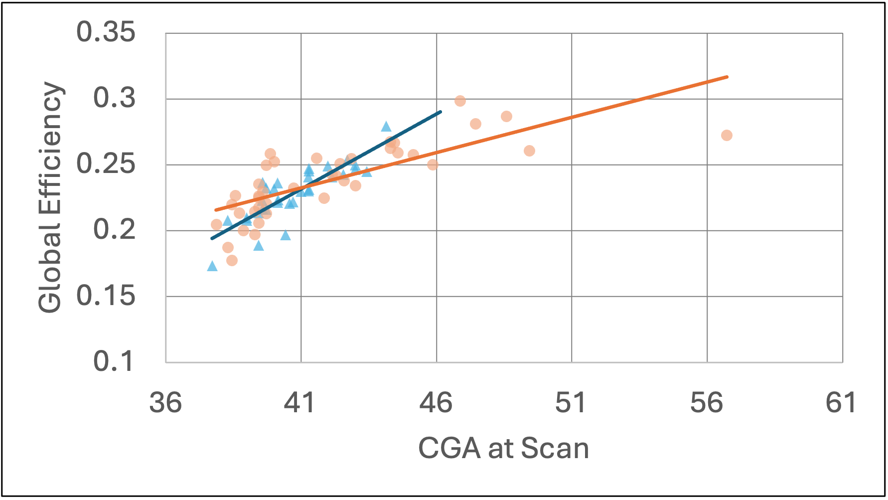
Plot includes global efficiency from pre- and post-operative brain MRIs for each subject with a best-fitted line. Infants with single ventricle physiology (SVP, orange line, N = 20) have a slower rate of development than those with d-Transposition of the Great Arteries (d-TGA, blue line, N = 16). Corrected gestational age (CGA).