352 - Critical early life events differently affect hippocampal development at term age: A comparison between preterm birth and neonatal hypoxic-ischemic encephalopathy
Friday, April 25, 2025
5:30pm - 7:45pm HST
Publication Number: 352.4173
Camille Heguy, McGill University Faculty of Medicine and Health Sciences, Montreal, PQ, Canada; Sarah Palmis, McGill University Faculty of Medicine and Health Sciences, Montreal, PQ, Canada; Bradley Karat, The University of Western Ontario - Schulich School of Medicine & Dentistry, London, ON, Canada; Christine Saint-Martin, McGill University Faculty of Medicine and Health Sciences, Montréal, PQ, Canada; Guillaume Gilbert, Philips Healthcare, Mississauga, ON, Canada; Olivier Fortin, Montreal Children's Hospital, Montreal, PQ, Canada; Emma G. Duerden, The University of Western Ontario - Schulich School of Medicine & Dentistry, London, ON, Canada; Pia Wintermark, McGill University, Montreal, PQ, Canada; Marie Brossard-Racine, McGill University Faculty of Medicine and Health Sciences, Montreal, PQ, Canada
Graduate student McGill University Faculty of Medicine and Health Sciences Montreal, Quebec, Canada
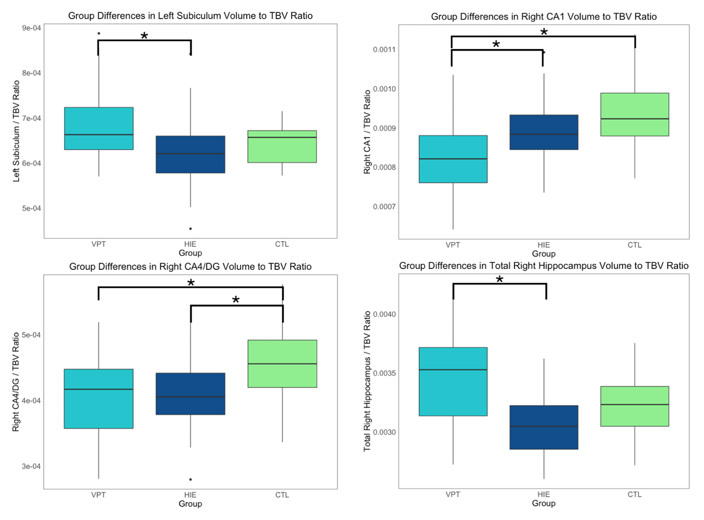
Background: The most active period of hippocampal growth occurs during the 2nd and 3rd trimesters of gestation and steadily continues up until the age of 2 years. Critical early life events [ELE], such as very preterm [VPT] birth ( < 32 weeks gestational age [GA]) or neonatal hypoxic-ischemic encephalopathy [HIE], coincide with this acute period of hippocampal growth, rendering this brain region vulnerable to developmental disruptions. Not surprisingly, previous studies have independently reported reduced hippocampal volumes in VPT and HIE individuals when compared to healthy peers during infancy and childhood. However, no study has directly compared regional hippocampal development between VPT and HIE neonates, which could clarify the specific developmental effects of these different ELE. Objective: To characterize and compare the distinct effects of 2 types of ELE on regional hippocampal development at term-equivalent age [TEA]. Design/Methods: As part of this study, we recruited VPT-born, term-born with moderate to severe HIE treated with therapeutic hypothermia, and healthy control [CTL] neonates without severe brain injury, congenital infection, or genetic anomaly. Enrollees completed a brain magnetic resonance imaging [MRI] at TEA. Total hippocampal volume and volumes of 7 subfields (subiculum, cornu ammonis [CA] 1 to 4, dentate gyrus [DG], and stratum radiatum, lacunosum, and moleculare) were extracted using Hippunfold, a validated automatic segmentation pipeline. Regional volume to total brain volume [TBV] ratios were calculated for each hippocampal region bilaterally and group differences were evaluated using ANOVA, correcting for multiple comparisons. Results: A total of 38 VPT, 28 HIE and 12 CTL neonates without severe brain injuries completed a brain MRI at a mean GA of 41 weeks. When compared to CTL, VPT neonates presented with significantly smaller right CA1 (p < 0.01) and CA4/DG (p < 0.05) ratios and HIE neonates presented with significantly smaller right CA4/DG ratios (p < 0.05), as well as smaller TBV (p < 0.05). When compared to HIE, VPT neonates presented with smaller right CA1 ratios (p < 0.05), greater left subiculum ratios (p < 0.05) and greater total right hippocampus ratios (p < 0.001).
Conclusion(s): Our results suggest that regional hippocampal development is differently affected by critical ELE. Although both VPT and HIE neonates showed increased vulnerability in the right CA4/DG region, premature extrauterine exposition had a more pronounced effect on right CA1 development at TEA compared to a term hypoxic-ischemic event. In contrast, HIE appears to negatively impact overall brain development.
Figure 1. Significant hippocampal ratio differences between VPT, HIE and CTL neonates.