337 - Novel Nomograms for Early Postnatal Brain Growth and Maturation in Extremely Preterm or Extremely Low Birth Weight Infants
Friday, April 25, 2025
5:30pm – 7:45pm HST
Publication Number: 337.5966
Lucia McLean, The Royal Women's Hospital, Carlton, Victoria, Australia; Brett J. Manley, Mercy Hospital for Women, Melbourne, Melbourne, Victoria, Australia; Louise S. Owen, Royal Women's Hospital, Melbourne, Melbourne, Victoria, Australia; Peter Davis, Royal Women's Hospital, Melbourne, Victoria, Australia; Rocco Cuzzilla, The Royal Women's Hospital, Parkville, Victoria, Australia
Neonatal Fellow The Royal Women's Hospital Carlton, Victoria, Australia
Background: Infants born extremely preterm or extremely low birth weight are at risk of neurodevelopmental impairment related to brain injury and aberrations in brain development. Simple and reliable linear measures made from neonatal neuroimaging have been shown to predict neurodevelopmental outcomes. Data on longitudinal measures of brain growth and maturation for extremely preterm infants are currently lacking, particularly for infants born < 25 weeks’ gestation. Objective: To describe brain growth and maturation for extremely preterm or extremely low birth weight infants using cranial ultrasonography (cUS) linear measures, and to develop nomograms from these longitudinal data. Design/Methods: Retrospective observational study of infants born < 28 weeks’ gestation or < 1000 g consecutively admitted to a single centre in 2021. Infants with congenital or genetic anomalies likely to affect brain growth or who died before admission or first cUS were excluded. Scans performed after development of major preterm brain injury were excluded. Simple and reliable linear measures (Figure 1) reflecting brain growth and maturation were made on cUS performed as part of clinical care. Imaging was routinely performed at, or around, postnatal days 1, 7-10, 42, and at term-equivalent age. Additional scans were performed as clinically indicated. Linear regression using mixed models were used to assess rates of change for each linear measure with respect to postnatal age (PNA) (weeks), adjusted for gestation. Nomograms were made for each linear measure of brain growth and maturation with respect to postmenstrual age (PMA) (weeks) and PNA (weeks). Results: 363 scans were assessed, including 101 scans for 28 infants born < 25 weeks’ gestation and 262 scans for 85 infants born ≥25 weeks’ gestation. The corpus callosum length, corpus callosum-fastigium distance, transcerebellar diameter, insular cortex height (left and right) and cingulate sulcus depth (left and right) increased, and the interopercular distance (left and right) decreased, with both PNA (Table) and PMA (not shown). Nomograms of linear measures were developed with respect to PNA and PMA (Figure 2).
Conclusion(s): Novel nomograms provide a reference for normative brain growth and maturation using simple and reliable cUS linear measures in extremely preterm or extremely low birth weight infants, including infants born < 25 weeks’ gestation.
Linear measures made on cranial ultrasonography 
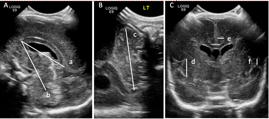
Cranial ultrasound linear measures of brain growth (A and B) and maturation (C). Images taken through the anterior fontanelle in the midsagittal plane (A) and coronal plane at the level of the foramina of Monro (C) and through the mastoid fontanelle in the coronal plane (B). a=corpus callosum length, b=corpus callosum-fastigium distance, c=transcerebellar diameter, d=insular cortex height, e=cingulate sulcus depth and f=interopercular opening.
Brain growth and maturation in extremely preterm or extremely low birth weight infants with respect to postnatal age
Transcerebellar diameter with respect to postnatal age and postmenstrual age