687 - Necrotizing Enterocolitis Pathogenesis Is Associated with Depletion of Tuft Cells in the Preterm Ileum
Monday, April 28, 2025
7:00am - 9:15am HST
Publication Number: 687.5876
Shirley Wang, OUHSC, Edmond, OK, United States; Hala Chaaban, Oklahoma University health sciences, Okl, OK, United States; Kathryn Burge, University of Oklahoma College of Medicine, Oklahoma City, OK, United States
Staff scentist OUHSC Edmond, Oklahoma, United States
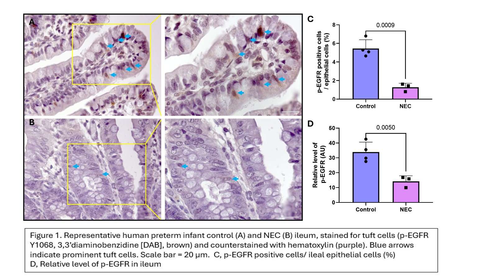
Background: Necrotizing enterocolitis (NEC) is a common and devastating intestinal emergency predominantly affecting preterm infants. Tuft cells (TCs), a rare, differentiated, epithelial chemosensory cell type, arise during the second trimester. Using apical ‘tufts’ of microfilaments, ileal TCs sense and respond to microbial signals, activating an intricate signal transduction pathway and driving cell fate decisions used to ‘sweep’ pathogens from the gut. In piglet models of human intestinal development, TC numbers peak at 25% of the epithelium at birth, a potential indication of their importance to the fetal/neonatal transition. However, ileal TC abundance in the human infant has been assumed to mirror the 1-2% prevalence of adult. Further, TCs are most prevalent and inducible in the distal ileum, the primary site of NEC mucosal injury. In inflammatory bowel disease, a condition with many similarities to NEC, TCs are lost during disease pathogenesis, while activation of TC signaling can prevent intestinal injury in animal models. The status of TCs during NEC pathogenesis, however, has not been explored. Objective: Identify functional markers for neonatal ileal TCs, and characterize TC abundance in the preterm infant, at homeostasis and during NEC pathogenesis Design/Methods: We performed immunohistochemical staining using a reported adult TC marker, phosphorylated epidermal growth factor receptor Y1068 (p-EGFR Y1068), to evaluate TC numbers and relative staining intensity in NEC (n = 3) and age-matched control (n = 4) ileum derived from preterm infant surgical resections Results: While cyclooxygenase-1, arachidonate 5-lipoxygenase, advillin, girdin, and choline acetyltransferase have been suggested as markers of human ileal TCs, p-EGFR proved an effective marker for human infant ileal TCs based on cell morphology. Interestingly, control TC abundance (Fig. 1A/C) was 5X that of NEC (Fig. 1B/C; p< 0.0009), and substantially elevated compared with adult. Further, p-EGFR staining intensity was significantly weaker in NEC compared with control tissue (Fig. 1D; p< 0.005), a further indication of the potential functional importance of p-EGFR to TC biology.
Conclusion(s): p-EGFR appears a suitable marker for evaluating TCs in the preterm infant epithelium, and the significant loss of ileal TCs in association with NEC pathogenesis implies a critical role for these chemosensory cells in regulating disease processes. Using this foundational knowledge, future experiments will attempt to determine whether loss of TCs is a cause or consequence of NEC pathophysiology.
p-EGFR-expressing tuft cells are reduced in NEC ileum