004 - Neuroprotective effect of therapeutic hypothermia after mild cerebral ischemia in term-equivalent fetal sheep
Monday, April 28, 2025
7:00am – 9:15am HST
Publication Number: 4.5808
Alice McDouall, The University of Auckland, Auckland, Auckland, New Zealand; Guido Wassink, University of auckland, Auckland, Auckland, New Zealand; Kelly Zhou, University of Auckland, Auckland, Auckland, New Zealand; Laura Bennet, The University of Auckland, Auckland, Auckland, New Zealand; Alistair J.. Gunn, The University of Auckland, Auckland, Auckland, New Zealand; Joanne Davidson, The University of Auckland, Auckland, Auckland, New Zealand
Associate Professor The University of Auckland Auckland, Auckland, New Zealand
Background: Infants with mild hypoxic ischemic encephalopathy (HIE) have increased risk of adverse neurological outcomes. These infants were largely excluded from the original clinical trials of therapeutic hypothermia, and so there are no approved treatment strategies for mild HIE. Despite this, there is therapeutic creep, with many infants with mild HIE now being treated with therapeutic hypothermia. Therefore, there is an urgent need to assess the impact of therapeutic hypothermia for mild hypoxic-ischemic (HI) brain injury. Objective: To determine whether therapeutic hypothermia is neuroprotective after mild cerebral ischemia in near-term fetal sheep. Design/Methods: Chronically instrumented fetal sheep at 0.85 gestation (term-equivalent) were exposed to sham-ischemia (n=8), mild ischemia induced by 10 minutes of bilateral carotid artery occlusion followed by normothermia (n=8) or mild ischemia with 72 h of hypothermia started 6 h after the end of ischemia (n=8). Sheep were killed after 7 days recovery for brain histology. Results: Ischemia was associated with reduced electroencephalogram (EEG) power in both the normothermia and hypothermia groups compared with sham controls, until 78 hours after ischemia (P < 0.05). In the normothermia group there was a significant decrease in spectral edge frequency after ischemia until 10 h, compared with sham control (P < 0.05). Hypothermia was associated with increased spectral edge frequency compared with sham controls and normothermia (P < 0.05). This was primarily mediated by an increase in the proportion of alpha activity compared with sham and normothermia groups (P < 0.05). Ischemia resulted in a significant loss of neurons within the CA1, CA3, CA4 and DG regions of the hippocampus (P < 0.05), while hypothermia significantly increased numbers of neurons in these regions (P < 0.05, Figure 1) to sham control levels.
Conclusion(s): These data suggest that therapeutic hypothermia for 72 hours is significantly neuroprotective when started 6 hours after the end of mild cerebral ischemia, with significantly improved neuronal survival in the hippocampus and increased high frequency alpha activity during cooling.
Numbers of NeuN-positive neurons in 4 regions of the hippocampus, 7 days after mild cerebral ischemia in the sham control, HI-normothermia and HI-hypothermia groups. 
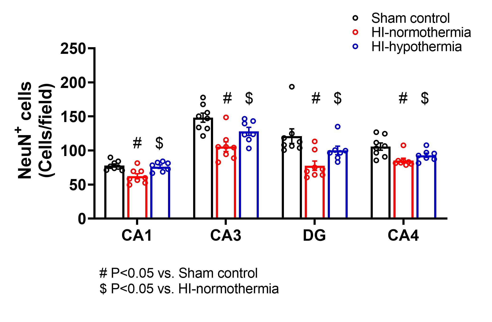
Ischemia was associated with a significant decrease in neuron numbers in all regions of the hippocampus, while therapeutic hypothermia significantly increased neuron number back to sham control level.