339 - Biometric Measurements of Corpus Callosum in Preterm and Term Infants
Friday, April 25, 2025
5:30pm – 7:45pm HST
Publication Number: 339.3933
Jordan P P. Cooper, University of South Alabama Children's and Women's Hospital, Mobile, AL, United States; Cristina Gavrilita, University of South Alabama Children's and Women's Hospital, Mobile, AL, United States; Michael Zayek, University of South Alabama, Mobile, AL, United States; Paul Maertens, University of South Alabama Children's and Women's Hospital, Mobile, AL, United States; Bianca M. Vamesu, University of South Alabama College of Medicine, Mobile, AL, United States
Pediatric Resident University of South Alabama Children's and Women's Hospital Mobile, Alabama, United States
Background: Corpus callosum (CC), the brain’s largest bundle of commissural fibers, serves as an important marker for assessing white matter development and injury. Abnormal CC size or impaired CC growth have been associated with adverse neurodevelopmental (ND) outcome. Objective: To establish and compare various CC biometric parameters (length, surface, and volume) measured shortly after birth using echoencephalogram. Design/Methods: Infants born at 22 0/7 to 40 0/7 weeks of gestation, admitted to our regional level III NICU between January 2017 and December 2023, and who had a cranial ultrasound on day 1 of life were enrolled. Exclusion criteria: multiple anomalies, brain anomalies, chromosomal anomalies, inborn errors of metabolism, chronic intrauterine infections, and abnormal cranial ultrasound.
A pediatric neurologist measured all CC parameters using midsagittal and coronal planes on the initial ultrasound. The length of CC was measured from the most anterior aspect of the genu to the most posterior aspect of the splenium (Fig.1A). The contour of the CC was manually delineated to determine its surface (Fig.1B). The volume of CC was calculated (product between the surface and the width of the CC) (Fig.1B, 1C). Results: CC measurements were obtained from 811 newborn infants by ultrasound within 24 hours after birth. While the growth of the CC length paralleled the head circumference growth from 22 weeks to 40 weeks of gestation, the CC surface area and volume doubled and tripled, respectively, during this developmental period (Table, Fig.2).
Conclusion(s): Thinning of the corpus callosum is frequently observed in extremely preterm infants. The measurements of CC surface area and volume may serve as more reliable predictors for ND impairment than CC length or head circumference. These normative values offer a reference for postnatal CC growth in preterm infants, facilitating early detection for developmental impairment risks.
Figure1 
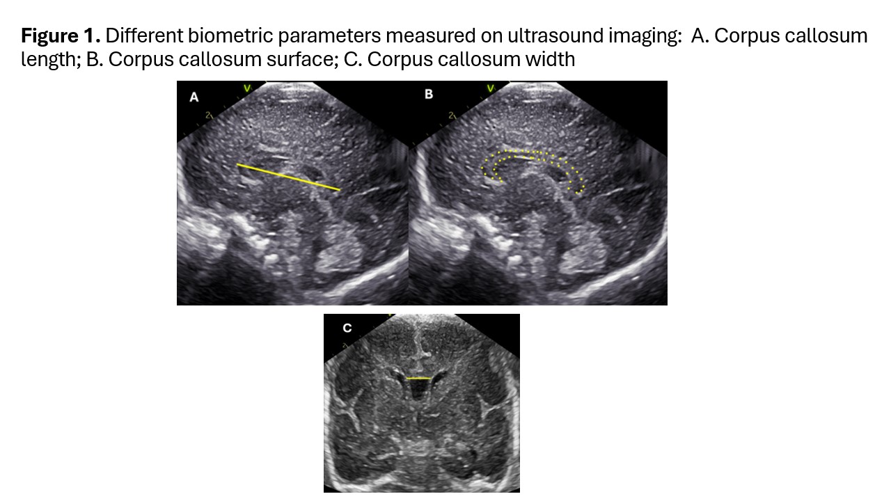
Corpus callosum measurements on ultrasound imaging
Table Biometric measurements of corpus callosum and head circumference by gestational age
Figure 2 Graphs showing length, surface area and volume of corpus callosum