
Neonatal General 5: Infectious Disease and Immunology
Session: Neonatal General 5: Infectious Disease and Immunology

Nickie Andescavage, MD
Associate Professor
Children's National Health System
Washington, District of Columbia, United States
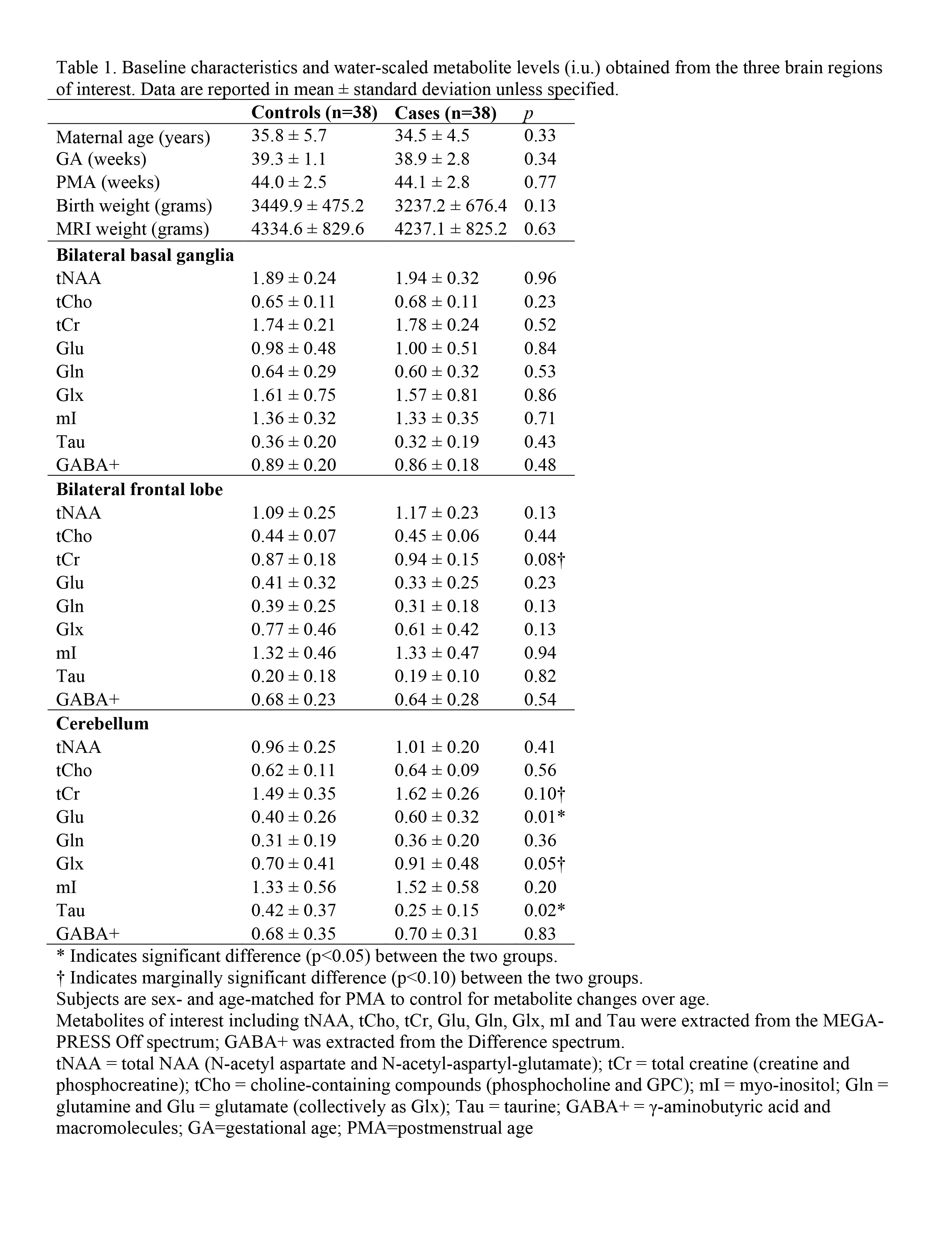 Baseline characteristics and water-scaled metabolite levels (i.u.) obtained from the three brain regions of interest. Data are reported in mean ± standard deviation unless specified.
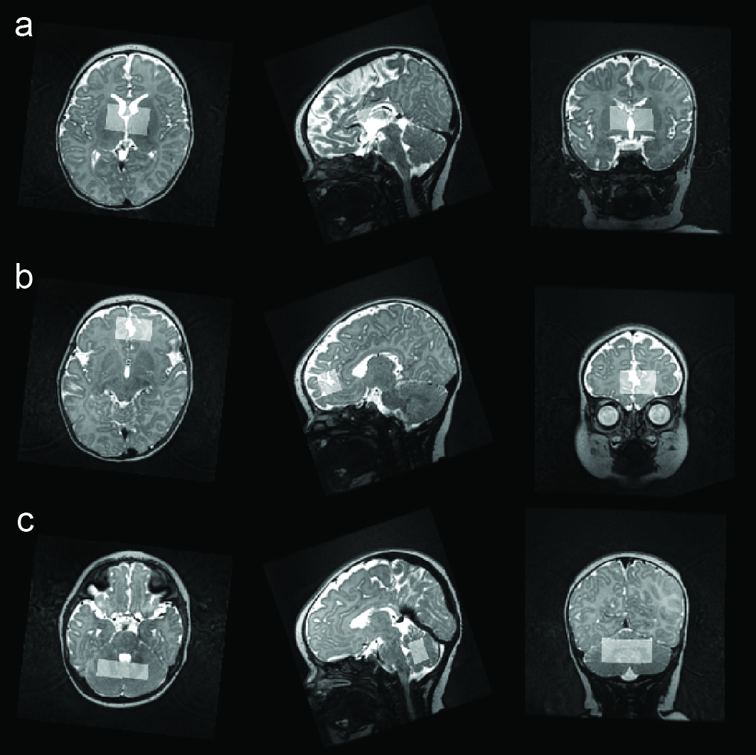 Representative voxel locations in (a) bilateral basal ganglia (38 x 16 x 22 mm^3), (b) bilateral frontal lobe (34 x 14 x 19 mm^3) and (c) cerebellum (31 x 11 x 16 mm^3) from a female neonate of mothers with COVID-19 exposure during pregnancy. Volume of the voxel was adjusted in each individual subject to optimize the placement and to avoid unwanted brain tissues.
.jpg) Boxplots for water-scaled metabolite levels (i.u.) in (a) the bilateral frontal lobe and (b) the cerebellum. In the bilateral frontal lobe, a noticeably difference (p=0.08) is observed in tCr between neonates of healthy mother (control) and neonates of mother with COVID-19 exposure during pregnancy (case). In the cerebellum, significant differences (p < 0.05) are observed in Glu and Tau between neonates of healthy mother (control) and neonates of mother with COVID-19 exposure during pregnancy (case). Of note, Glx is marginal significantly different (p=0.05) between the two groups driven by the increase of Glu. tCr also has a strong increasing trend in the case group.
Boxplots for water-scaled metabolite levels (i.u.) in (a) the bilateral frontal lobe and (b) the cerebellum. In the bilateral frontal lobe, a noticeably difference (p=0.08) is observed in tCr between neonates of healthy mother (control) and neonates of mother with COVID-19 exposure during pregnancy (case). In the cerebellum, significant differences (p < 0.05) are observed in Glu and Tau between neonates of healthy mother (control) and neonates of mother with COVID-19 exposure during pregnancy (case). Of note, Glx is marginal significantly different (p=0.05) between the two groups driven by the increase of Glu. tCr also has a strong increasing trend in the case group.